当前位置:
X-MOL 学术
›
J. Raman Spectrosc.
›
论文详情
Our official English website, www.x-mol.net, welcomes your feedback! (Note: you will need to create a separate account there.)
Raman microspectroscopy for label‐free diagnosis of amyloid light‐chain amyloidosis in various organs
Journal of Raman Spectroscopy ( IF 2.5 ) Pub Date : 2024-03-19 , DOI: 10.1002/jrs.6665 Shin‐ichiro Yanagiya 1, 2 , Takeshi Honda 2 , Hiroki Takanari 1, 2, 3 , Kimiko Sogabe 4 , Shingen Nakamura 5 , Yoshimi Bando 6 , Koichi Tsuneyama 7 , Masahiro Abe 8 , Hirokazu Miki 9
Journal of Raman Spectroscopy ( IF 2.5 ) Pub Date : 2024-03-19 , DOI: 10.1002/jrs.6665 Shin‐ichiro Yanagiya 1, 2 , Takeshi Honda 2 , Hiroki Takanari 1, 2, 3 , Kimiko Sogabe 4 , Shingen Nakamura 5 , Yoshimi Bando 6 , Koichi Tsuneyama 7 , Masahiro Abe 8 , Hirokazu Miki 9
Affiliation
|
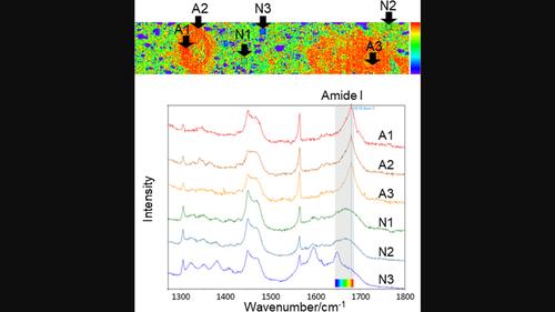
Systemic amyloidosis is a group of diseases in which misfolded proteins aggregate as fibrous amyloid proteins with a β‐sheet structure and deposit in organs, resulting in organ failure. Most types of amyloidosis have a poor prognosis, and prompt diagnosis is essential for treatment. Systemic immunoglobulin light‐chain (AL) amyloidosis is a type of amyloidosis that occurs when abnormal immunoglobulin light‐chain proteins are deposited in various organs and tissues. The deposition of amyloid proteins in tissues has traditionally been confirmed using Congo red staining and polarised light microscopy, which show apple‐green birefringence. In this study, we aimed to verify whether amyloid deposition in the heart, kidney, rectum, duodenum and skin can be detected using Raman microspectroscopy. Serial sections were prepared from formalin‐fixed paraffin‐embedded tissue biopsy samples obtained from patients with systemic amyloidosis. One of the serial sections was stained with Congo red to confirm the deposition of amyloid proteins using polarised light microscopy, whereas the other was left unstained for Raman microspectroscopy. A characteristic peak at Raman shift of 1665–1680 cm−1 , which may represent a β‐sheet structure of amyloid proteins, was recorded in the area where the amyloid deposition had been confirmed by Congo red staining. Based on the peak at 1640–1680 cm−1 , a colour map was obtained to detect amyloid protein‐positive regions. Thus, amyloid protein detection using Raman microspectroscopy may be useful for rapid diagnosis of amyloidosis.
中文翻译:

拉曼显微光谱法用于无标记诊断各种器官中淀粉样蛋白轻链淀粉样变性
系统性淀粉样变性是一组错误折叠的蛋白质聚集成具有β折叠结构的纤维状淀粉样蛋白并沉积在器官中,导致器官衰竭的疾病。大多数类型的淀粉样变性预后不良,及时诊断对于治疗至关重要。系统性免疫球蛋白轻链(AL)淀粉样变性是一种淀粉样变性,当异常的免疫球蛋白轻链蛋白沉积在各个器官和组织中时发生。传统上,淀粉样蛋白在组织中的沉积是通过刚果红染色和偏光显微镜来证实的,偏光显微镜显示出苹果绿双折射。在本研究中,我们旨在验证是否可以使用拉曼显微光谱检测心脏、肾脏、直肠、十二指肠和皮肤中的淀粉样蛋白沉积。使用从系统性淀粉样变性患者获得的福尔马林固定石蜡包埋的组织活检样本制备连续切片。其中一个连续切片用刚果红染色,以使用偏光显微镜确认淀粉样蛋白的沉积,而另一个切片则未染色,用于拉曼显微光谱学。拉曼位移 1665–1680 cm 处的特征峰−1 ,可能代表淀粉样蛋白的β-折叠结构,记录在通过刚果红染色证实淀粉样蛋白沉积的区域。基于 1640–1680 cm 处的峰值−1 ,获得彩色图来检测淀粉样蛋白阳性区域。因此,使用拉曼显微光谱检测淀粉样蛋白可能有助于淀粉样变性的快速诊断。
更新日期:2024-03-19
中文翻译:
拉曼显微光谱法用于无标记诊断各种器官中淀粉样蛋白轻链淀粉样变性
系统性淀粉样变性是一组错误折叠的蛋白质聚集成具有β折叠结构的纤维状淀粉样蛋白并沉积在器官中,导致器官衰竭的疾病。大多数类型的淀粉样变性预后不良,及时诊断对于治疗至关重要。系统性免疫球蛋白轻链(AL)淀粉样变性是一种淀粉样变性,当异常的免疫球蛋白轻链蛋白沉积在各个器官和组织中时发生。传统上,淀粉样蛋白在组织中的沉积是通过刚果红染色和偏光显微镜来证实的,偏光显微镜显示出苹果绿双折射。在本研究中,我们旨在验证是否可以使用拉曼显微光谱检测心脏、肾脏、直肠、十二指肠和皮肤中的淀粉样蛋白沉积。使用从系统性淀粉样变性患者获得的福尔马林固定石蜡包埋的组织活检样本制备连续切片。其中一个连续切片用刚果红染色,以使用偏光显微镜确认淀粉样蛋白的沉积,而另一个切片则未染色,用于拉曼显微光谱学。拉曼位移 1665–1680 cm 处的特征峰


 京公网安备 11010802027423号