Functional & Integrative Genomics ( IF 2.9 ) Pub Date : 2024-03-20 , DOI: 10.1007/s10142-024-01324-z Zeinab Moradi , Mandana Kazemi , Roya Jamshidi-Khalifelou , Vahid Bahramnia , Fatemeh Esfandmaz , Reza Rahnavard , Behnoush Moradgholi , Tohid Piri-Gharaghie
|
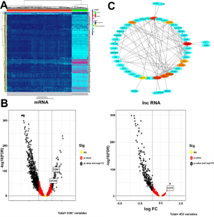
This research provides a glimmer of hope that the knockout of HCP5 leads to a therapy response to considerably prolong the life of patients with OC. RT-PCR evaluated the expression of lncRNA HCP5 in the ovarian cancer OVCAR-3 cell line. CRISPR knockout cell lines validated by western blot. Small genomic deletions at the targeted locus were induced. CCK-8 colony formation assays were used to analyze the effect of HCP5 knockout on the proliferation capacity of OVCAR-3 cells. Transwell migration and invasion assayed. Furthermore, the Sphere-formation assay isolated the most aggressive population of cancer stem cells. Bioinformatic analysis showed a significant correlation between lncRNA HCP5 up-regulation and OVCAR-3 cell proliferation. The ChIP technique assesses specific sites of interaction between transcription factors and DNA. Real-time PCR assays explored the relationship between HCP5, Hsa-miR-9-5p, CXCR4, CDH1, caspase-3, p53, bcl2 and survivin. PCR carried out amplification of the 448-bp band for sgRNA1 and sgRNA2 after the use of particular primers for HCP5. the number of breast cancer cells that moved to the bottom chamber reduced considerably after transfection with PX461-sgRNA1/2 vectors compared to the Blank control groups (P < 0.05). MTT assay designated growth curves that showed the rate of OVCAR-3 growth was significantly repressed (***P < 0.001) when compared with control OVCAR-3 cells after HCP5 knockdown. Also, the survival results of W.T cells in 24, 48 and 72 h showed 92%, 87% and 85%, respectively. This is while the cells of the CRISPR/Cas9 group in which LncRNA HCP5 was knocked out had 42% (*P < 0.05), 23%(**P < 0.01) and 14% (**P < 0.01) survival, respectively. The expression levels of caspase-3, Hsa-miR-9-5p, P53 genes in the HCP5 deletion of CRISPR/Cas9 group significantly increased than the W.T. control group; the deletion group showed a considerable reduction in HCP5 expression compared to the blank control group (3.6-fold, p < 0.01). Whereas BCL2, SURVIVIN, CXCR4, CDH1 genes expression markedly increased than in HCP5 knockout cells (5.8-fold, p < 0.05). These results indicate that CRISPR/Cas9‐mediated HCP5 disruption on OVCAR-3 cell lines promotes anti‐tumor biomarkers, suppressing ovarian cancer progression. Consistent with these results, HCP5 is one of the most critical lnc for the efficient proliferation and migration of OVCAR-3 cell lines.
中文翻译:
CRISPR du-HITI 是一种靶向长非编码 RNA HCP5 作为卵巢癌细胞增殖抑制因子的有吸引力的方法
这项研究带来了一线希望,即敲除 HCP5 会产生治疗反应,从而显着延长 OC 患者的生命。 RT-PCR 评估了卵巢癌 OVCAR-3 细胞系中 lncRNA HCP5 的表达。通过蛋白质印迹验证 CRISPR 敲除细胞系。诱导了目标位点的小基因组缺失。采用CCK-8集落形成实验分析HCP5敲除对OVCAR-3细胞增殖能力的影响。检测 Transwell 迁移和侵袭。此外,球体形成测定分离出了最具攻击性的癌症干细胞群。生物信息分析显示lncRNA HCP5上调与OVCAR-3细胞增殖之间存在显着相关性。 ChIP 技术评估转录因子和 DNA 之间相互作用的特定位点。实时 PCR 检测探讨了 HCP5、Hsa-miR-9-5p、CXCR4、CDH1、caspase-3、p53、bcl2 和生存素之间的关系。在使用 HCP5 的特定引物后,PCR 对 sgRNA1 和 sgRNA2 的 448 bp 条带进行了扩增。转染 PX461-sgRNA1/2 载体后,与空白对照组相比,移至底室的乳腺癌细胞数量明显减少(P < 0.05)。 MTT 测定指定的生长曲线显示, 与 HCP5 敲低后的对照 OVCAR-3 细胞相比,OVCAR-3生长速率显着受到抑制 (*** P < 0.001)。此外,WT细胞在24、48和72小时的存活率分别为92%、87%和85%。而敲除 LncRNA HCP5 的 CRISPR/Cas9 组细胞的 存活率分别为 42% (* P < 0.05)、23% (** P < 0.01) 和 14% (** P < 0.01)。 。 CRISPR/Cas9 HCP5缺失组中caspase-3、Hsa-miR-9-5p、P53基因表达量较WT对照组显着升高;与空白对照组相比,缺失组的 HCP5 表达显着降低(3.6 倍,p < 0.01)。而 BCL2、SURVIVIN、CXCR4、CDH1 基因表达比 HCP5 敲除细胞中显着增加(5.8 倍,p < 0.05)。这些结果表明 CRISPR/Cas9 介导的 OVCAR-3 细胞系上的 HCP5 破坏可促进抗肿瘤生物标志物的产生,从而抑制卵巢癌的进展。与这些结果一致,HCP5是OVCAR-3细胞系有效增殖和迁移的最关键的lnc之一。


 京公网安备 11010802027423号