Journal of Neuroimmune Pharmacology ( IF 6.2 ) Pub Date : 2024-03-26 , DOI: 10.1007/s11481-024-10108-y Hung-Chuan Pan , Cheng-Ning Yang , Wen-Jane Lee , Jason Sheehan , Sheng-Mao Wu , Hong-Shiu Chen , Mao-Hsun Lin , Li-Wei Shen , Shu-Hua Lee , Chin-Chang Shen , Liang-Yi Pan , Shing‑Hwa Liu , Meei-Ling Sheu
|
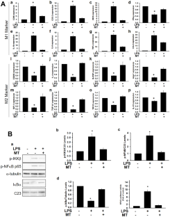
Neuro-inflammation involves distinct alterations of microglial phenotypes, containing nocuous pro-inflammatory M1-phenotype and neuroprotective anti-inflammatory M-phenotype. Currently, there is no effective treatment for modulating such alterations. M1/M2 marker of primary microglia influenced by Melatonin were detected via qPCR. Functional activities were explored by western blotting, luciferase activity, EMSA, and ChIP assay. Structure interaction was assessed by molecular docking and LIGPLOT analysis. ER-stress detection was examined by ultrastructure TEM, calapin activity, and ERSE assay. The functional neurobehavioral evaluations were used for investigation of Melatonin on the neuroinflammation in vivo. Melatonin had targeted on Peroxisome Proliferator Activated Receptor Delta (PPARδ) activity, boosted LPS-stimulated alterations in polarization from the M1 to the M2 phenotype, and thereby inhibited NFκB–IKKβ activation in primary microglia. The PPARδ agonist L-165,041 or over-expression of PPARδ plasmid (ov-PPARδ) showed similar results. Molecular docking screening, dynamic simulation approaches, and biological studies of Melatonin showed that the activated site was located at PPARδ (phospho-Thr256-PPARδ). Activated microglia had lowered PPARδ activity as well as the downstream SIRT1 formation via enhancing ER-stress. Melatonin, PPARδ agonist and ov-PPARδ all effectively reversed the above-mentioned effects. Melatonin blocked ER-stress by regulating calapin activity and expression in LPS-activated microglia. Additionally, Melatonin or L-165,041 ameliorated the neurobehavioral deficits in LPS-aggravated neuroinflammatory mice through blocking microglia activities, and also promoted phenotype changes to M2-predominant microglia. Melatonin suppressed neuro-inflammation in vitro and in vivo by tuning microglial activation through the ER-stress-dependent PPARδ/SIRT1 signaling cascade. This treatment strategy is an encouraging pharmacological approach for the remedy of neuro-inflammation associated disorders.
Graphical Abstract
Schematic of Proposed Mechanism for the role of Melatonin in Activated Microglia and neuro-inflammation Effects. LPS-induced ER Stress and Regulated PPARδ Expression, NFκB Phosphorylation, Subsequently Reduces SIRT1 Expression and then Triggers the Microglia Activation and Brain Damage. In the Present Study, we Provide the Evidence to Demonstrate that Melatonin Plays a Potential Protective role in Neuroprotective Effects through PPARδ/SIRT1 Pathway. In Addition, PPARδ Pharmacological Agonists L165041 also Possessed Similar Effects. These Results Suggest that the Activation of PPARδ/SIRT1 by Melatonin could Counteract the Detrimental Effect of LPS. Also, the Results Suggest Melatonin may Exert a Therapeutic Effect for Neuroinflammatory Disorders
中文翻译:
褪黑激素通过调节 ER 应激/PPARδ/SIRT1 信号轴增强神经炎症大鼠模型中小胶质细胞 M2 极化
神经炎症涉及小胶质细胞表型的明显改变,包括有害的促炎 M1 表型和神经保护性抗炎 M 表型。目前,没有有效的治疗方法来调节这种改变。通过 qPCR 检测受褪黑素影响的原代小胶质细胞的 M1/M2 标记。通过蛋白质印迹、荧光素酶活性、EMSA 和 ChIP 测定来探索功能活性。通过分子对接和 LIGPLOT 分析评估结构相互作用。通过超微结构 TEM、calapin 活性和 ERSE 测定检查 ER 应激检测。功能性神经行为评估用于研究褪黑素对体内神经炎症的影响。褪黑素靶向过氧化物酶体增殖物激活受体 Delta (PPARδ) 活性,促进 LPS 刺激的从 M1 表型到 M2 表型的极化改变,从而抑制原代小胶质细胞中 NFκB-IKKβ 的激活。 PPARδ 激动剂 L-165,041 或 PPARδ 质粒(ov-PPARδ)的过表达显示出类似的结果。褪黑素的分子对接筛选、动态模拟方法和生物学研究表明,其激活位点位于PPARδ(磷酸化Thr256-PPARδ)。激活的小胶质细胞通过增强 ER 应激降低了 PPARδ 活性以及下游 SIRT1 的形成。褪黑激素、PPARδ激动剂和ov-PPARδ均能有效逆转上述效应。褪黑激素通过调节 LPS 激活的小胶质细胞中的 calapin 活性和表达来阻断 ER 应激。此外,褪黑素或 L-165,041 通过阻断小胶质细胞活动改善 LPS 加重的神经炎症小鼠的神经行为缺陷,并促进 M2 为主的小胶质细胞的表型变化。褪黑素通过 ER 应激依赖性 PPARδ/SIRT1 信号级联调节小胶质细胞的激活,在体外和体内抑制神经炎症。这种治疗策略是治疗神经炎症相关疾病的令人鼓舞的药理学方法。
图形概要
褪黑激素在激活的小胶质细胞和神经炎症效应中的作用的拟议机制示意图。 LPS 诱导 ER 应激并调节 PPARδ 表达、NFκB 磷酸化,随后降低 SIRT1 表达,进而触发小胶质细胞激活和脑损伤。在本研究中,我们提供证据来证明褪黑激素通过 PPARδ/SIRT1 途径在神经保护作用中发挥潜在的保护作用。此外,PPARδ药理激动剂L165041也具有类似的作用。这些结果表明,褪黑激素激活 PPARδ/SIRT1 可以抵消 LPS 的有害影响。此外,结果表明褪黑激素可能对神经炎症性疾病有治疗作用


 京公网安备 11010802027423号