当前位置:
X-MOL 学术
›
Comput. Struct. Biotechnol. J.
›
论文详情
Our official English website, www.x-mol.net, welcomes your feedback! (Note: you will need to create a separate account there.)
Classifying breast cancer and fibroadenoma tissue biopsies from paraffined stain-free slides by fractal biomarkers in Fourier Ptychographic Microscopy
Computational and Structural Biotechnology Journal ( IF 6 ) Pub Date : 2024-03-24 , DOI: 10.1016/j.csbj.2024.03.019 Vittorio Bianco , Marika Valentino , Daniele Pirone , Lisa Miccio , Pasquale Memmolo , Valentina Brancato , Luigi Coppola , Giovanni Smaldone , Massimiliano D’Aiuto , Gennaro Mossetti , Marco Salvatore , Pietro Ferraro
Computational and Structural Biotechnology Journal ( IF 6 ) Pub Date : 2024-03-24 , DOI: 10.1016/j.csbj.2024.03.019 Vittorio Bianco , Marika Valentino , Daniele Pirone , Lisa Miccio , Pasquale Memmolo , Valentina Brancato , Luigi Coppola , Giovanni Smaldone , Massimiliano D’Aiuto , Gennaro Mossetti , Marco Salvatore , Pietro Ferraro
|
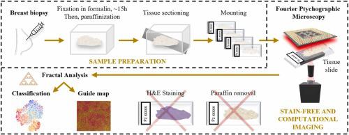
Breast cancer is one of the most spread and monitored pathologies in high-income countries. After breast biopsy, histological tissue is stored in paraffin, sectioned and mounted. Conventional inspection of tissue slides under benchtop light microscopes involves paraffin removal and staining, typically with H&E. Then, expert pathologists are called to judge the stained slides. However, paraffin removal and staining are operator-dependent, time and resources consuming processes that can generate ambiguities due to non-uniform staining. Here we propose a novel method that can work directly on paraffined stain-free slides. We use Fourier Ptychography as a quantitative phase-contrast microscopy method, which allows accessing a very wide field of view (i.e., mm) in one single image while guaranteeing high lateral resolution (i.e., 0.5 µm). This imaging method is multi-scale, since it enables looking at the big picture, i.e. the complex tissue structure and connections, with the possibility to zoom-in up to the single-cell level. To handle this informative image content, we introduce elements of fractal geometry as multi-scale analysis method. We show the effectiveness of fractal features in describing and classifying fibroadenoma and breast cancer tissue slides from ten patients with very high accuracy. We reach 94.0 ± 4.2% test accuracy in classifying single images. Above all, we show that combining the decisions of the single images, each patient’s slide can be classified with no error. Besides, fractal geometry returns a guide map to help pathologist to judge the different tissue portions based on the likelihood these can be associated to a breast cancer or fibroadenoma biomarker. The proposed automatic method could significantly simplify the steps of tissue analysis and make it independent from the sample preparation, the skills of the lab operator and the pathologist.
中文翻译:

通过傅里叶叠层显微镜中的分形生物标志物对来自石蜡无染色载玻片的乳腺癌和纤维腺瘤组织活检进行分类
乳腺癌是高收入国家中传播最广泛、监测最严重的疾病之一。乳房活检后,将组织学组织保存在石蜡中,切片并封片。在台式光学显微镜下对组织载玻片进行的常规检查涉及石蜡去除和染色,通常使用 H&E。然后,专家病理学家被要求对染色的载玻片进行判断。然而,石蜡去除和染色是依赖于操作员的、耗时和资源消耗的过程,可能由于不均匀的染色而产生歧义。在这里,我们提出了一种可以直接在石蜡无污玻片上工作的新颖方法。我们使用傅里叶叠层成像术作为定量相差显微镜方法,它允许在单个图像中获得非常宽的视场(即毫米),同时保证高横向分辨率(即0.5微米)。这种成像方法是多尺度的,因为它能够观察大图景,即复杂的组织结构和连接,并且可以放大到单细胞水平。为了处理这些信息丰富的图像内容,我们引入分形几何元素作为多尺度分析方法。我们展示了分形特征在描述和分类来自 10 名患者的纤维腺瘤和乳腺癌组织切片方面的有效性,并且具有非常高的准确性。我们在对单幅图像进行分类时达到了 94.0 ± 4.2% 的测试准确率。最重要的是,我们表明,结合单个图像的决策,每个患者的幻灯片都可以毫无错误地进行分类。此外,分形几何返回指导图,帮助病理学家根据这些组织部分与乳腺癌或纤维腺瘤生物标志物相关的可能性来判断不同的组织部分。所提出的自动方法可以显着简化组织分析的步骤,并使其独立于样品制备、实验室操作员和病理学家的技能。
更新日期:2024-03-24
中文翻译:
通过傅里叶叠层显微镜中的分形生物标志物对来自石蜡无染色载玻片的乳腺癌和纤维腺瘤组织活检进行分类
乳腺癌是高收入国家中传播最广泛、监测最严重的疾病之一。乳房活检后,将组织学组织保存在石蜡中,切片并封片。在台式光学显微镜下对组织载玻片进行的常规检查涉及石蜡去除和染色,通常使用 H&E。然后,专家病理学家被要求对染色的载玻片进行判断。然而,石蜡去除和染色是依赖于操作员的、耗时和资源消耗的过程,可能由于不均匀的染色而产生歧义。在这里,我们提出了一种可以直接在石蜡无污玻片上工作的新颖方法。我们使用傅里叶叠层成像术作为定量相差显微镜方法,它允许在单个图像中获得非常宽的视场(即毫米),同时保证高横向分辨率(即0.5微米)。这种成像方法是多尺度的,因为它能够观察大图景,即复杂的组织结构和连接,并且可以放大到单细胞水平。为了处理这些信息丰富的图像内容,我们引入分形几何元素作为多尺度分析方法。我们展示了分形特征在描述和分类来自 10 名患者的纤维腺瘤和乳腺癌组织切片方面的有效性,并且具有非常高的准确性。我们在对单幅图像进行分类时达到了 94.0 ± 4.2% 的测试准确率。最重要的是,我们表明,结合单个图像的决策,每个患者的幻灯片都可以毫无错误地进行分类。此外,分形几何返回指导图,帮助病理学家根据这些组织部分与乳腺癌或纤维腺瘤生物标志物相关的可能性来判断不同的组织部分。所提出的自动方法可以显着简化组织分析的步骤,并使其独立于样品制备、实验室操作员和病理学家的技能。


 京公网安备 11010802027423号