当前位置:
X-MOL 学术
›
WIREs Nanomed. Nanobiotechnol.
›
论文详情
Our official English website, www.x-mol.net, welcomes your feedback! (Note: you will need to create a separate account there.)
Using imaging modalities to predict nanoparticle distribution and treatment efficacy in solid tumors: The growing role of ultrasound
WIREs Nanomedicine and Nanobiotechnology ( IF 8.6 ) Pub Date : 2024-04-01 , DOI: 10.1002/wnan.1957 Michaela B. Cooley 1 , Dana Wegierak 1 , Agata A. Exner 1, 2
WIREs Nanomedicine and Nanobiotechnology ( IF 8.6 ) Pub Date : 2024-04-01 , DOI: 10.1002/wnan.1957 Michaela B. Cooley 1 , Dana Wegierak 1 , Agata A. Exner 1, 2
Affiliation
|
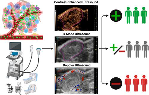
Nanomedicine in oncology has not had the success in clinical impact that was anticipated in the early stages of the field's development. Ideally, nanomedicines selectively accumulate in tumor tissue and reduce systemic side effects compared to traditional chemotherapeutics. However, this has been more successful in preclinical animal models than in humans. The causes of this failure to translate may be related to the intra- and inter-patient heterogeneity of the tumor microenvironment. Predicting whether a patient will respond positively to treatment prior to its initiation, through evaluation of characteristics like nanoparticle extravasation and retention potential in the tumor, may be a way to improve nanomedicine success rate. While there are many potential strategies to accomplish this, prediction and patient stratification via noninvasive medical imaging may be the most efficient and specific strategy. There have been some preclinical and clinical advances in this area using MRI, CT, PET, and other modalities. An alternative approach that has not been studied as extensively is biomedical ultrasound, including techniques such as multiparametric contrast-enhanced ultrasound (mpCEUS), doppler, elastography, and super-resolution processing. Ultrasound is safe, inexpensive, noninvasive, and capable of imaging the entire tumor with high temporal and spatial resolution. In this work, we summarize the in vivo imaging tools that have been used to predict nanoparticle distribution and treatment efficacy in oncology. We emphasize ultrasound imaging and the recent developments in the field concerning CEUS. The successful implementation of an imaging strategy for prediction of nanoparticle accumulation in tumors could lead to increased clinical translation of nanomedicines, and subsequently, improved patient outcomes.
中文翻译:

使用成像方式预测实体瘤中纳米颗粒的分布和治疗效果:超声的作用日益增强
肿瘤学纳米医学在临床影响方面尚未取得该领域发展早期阶段预期的成功。理想情况下,与传统化疗药物相比,纳米药物选择性地积聚在肿瘤组织中并减少全身副作用。然而,这在临床前动物模型中比在人类中更成功。这种翻译失败的原因可能与患者内和患者间肿瘤微环境的异质性有关。通过评估纳米颗粒外渗和肿瘤中保留潜力等特征,预测患者在治疗开始前是否会对治疗产生积极反应,可能是提高纳米医学成功率的一种方法。虽然有许多潜在的策略可以实现这一目标,但通过无创医学成像进行预测和患者分层可能是最有效和最具体的策略。利用 MRI、CT、PET 和其他方式在该领域取得了一些临床前和临床进展。另一种尚未得到广泛研究的替代方法是生物医学超声,包括多参数对比增强超声 (mpCEUS)、多普勒、弹性成像和超分辨率处理等技术。超声安全、廉价、无创,并且能够以高时间和空间分辨率对整个肿瘤进行成像。在这项工作中,我们总结了用于预测肿瘤学中纳米颗粒分布和治疗效果的体内成像工具。我们重点关注超声成像以及 CEUS 领域的最新发展。成功实施预测肿瘤中纳米颗粒积累的成像策略可能会增加纳米药物的临床转化,从而改善患者的治疗结果。
更新日期:2024-04-02
中文翻译:
使用成像方式预测实体瘤中纳米颗粒的分布和治疗效果:超声的作用日益增强
肿瘤学纳米医学在临床影响方面尚未取得该领域发展早期阶段预期的成功。理想情况下,与传统化疗药物相比,纳米药物选择性地积聚在肿瘤组织中并减少全身副作用。然而,这在临床前动物模型中比在人类中更成功。这种翻译失败的原因可能与患者内和患者间肿瘤微环境的异质性有关。通过评估纳米颗粒外渗和肿瘤中保留潜力等特征,预测患者在治疗开始前是否会对治疗产生积极反应,可能是提高纳米医学成功率的一种方法。虽然有许多潜在的策略可以实现这一目标,但通过无创医学成像进行预测和患者分层可能是最有效和最具体的策略。利用 MRI、CT、PET 和其他方式在该领域取得了一些临床前和临床进展。另一种尚未得到广泛研究的替代方法是生物医学超声,包括多参数对比增强超声 (mpCEUS)、多普勒、弹性成像和超分辨率处理等技术。超声安全、廉价、无创,并且能够以高时间和空间分辨率对整个肿瘤进行成像。在这项工作中,我们总结了用于预测肿瘤学中纳米颗粒分布和治疗效果的体内成像工具。我们重点关注超声成像以及 CEUS 领域的最新发展。成功实施预测肿瘤中纳米颗粒积累的成像策略可能会增加纳米药物的临床转化,从而改善患者的治疗结果。


 京公网安备 11010802027423号