Our official English website, www.x-mol.net, welcomes your feedback! (Note: you will need to create a separate account there.)
Morphological and electrophysiological characterization of a novel displaced astrocyte in the mouse retina
Glia ( IF 6.2 ) Pub Date : 2024-04-09 , DOI: 10.1002/glia.24536 Joseph Matthew Holden 1, 2 , Lauren Katie Wareham 1 , David John Calkins 1
Glia ( IF 6.2 ) Pub Date : 2024-04-09 , DOI: 10.1002/glia.24536 Joseph Matthew Holden 1, 2 , Lauren Katie Wareham 1 , David John Calkins 1
Affiliation
|
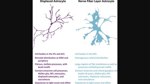
Astrocytes throughout the central nervous system are heterogeneous in both structure and function. This diversity leads to tissue‐specific specialization where morphology is adapted to the surrounding neuronal circuitry, as seen in Bergman glia of the cerebellum and Müller glia of the retina. Because morphology can be a differentiating factor for cellular classification, we recently developed a mouse where glial‐fibrillary acidic protein (GFAP)‐expressing cells stochastically label for full membranous morphology. Here we utilize this tool to investigate whether morphological and electrophysiological features separate types of mouse retinal astrocytes. In this work, we report on a novel glial population found in the inner plexiform layer and ganglion cell layer which expresses the canonical astrocyte markers GFAP, S100β, connexin‐43, Sox2 and Sox9. Apart from their retinal layer localization, these cells are unique in their radial distribution. They are notably absent from the mid‐retina but are heavily concentrated near the optic nerve head, and to a lesser degree the peripheral retina. Additionally, their morphology is distinct from both nerve fiber layer astrocytes and Müller glia, appearing more similar to amacrine cells. Despite this structural similarity, these cells lack protein expression of common neuronal markers. Additionally, they do not exhibit action potentials, but rather resemble astrocytes and Müller glia in their small amplitude, graded depolarization to both light onset and offset. Their structure, protein expression, physiology, and intercellular connections suggest that these cells are astrocytic, displaced from their counterparts in the nerve fiber layer. As such, we refer to these cells as displaced retinal astrocytes.
中文翻译:

小鼠视网膜中新型移位星形胶质细胞的形态学和电生理学特征
整个中枢神经系统的星形胶质细胞在结构和功能上都是异质的。这种多样性导致了组织特异性的特化,其中形态适应周围的神经元回路,如小脑的伯格曼神经胶质细胞和视网膜的穆勒神经胶质细胞所示。由于形态可能是细胞分类的区分因素,因此我们最近开发了一种小鼠,其中表达胶质纤维酸性蛋白(GFAP)的细胞随机标记全膜形态。在这里,我们利用这个工具来研究形态和电生理特征是否区分小鼠视网膜星形胶质细胞的类型。在这项工作中,我们报告了在内丛状层和神经节细胞层中发现的一种新型神经胶质细胞群,其表达经典星形胶质细胞标记物 GFAP、S100β、connexin-43、Sox2 和 Sox9。除了视网膜层定位之外,这些细胞的径向分布也是独特的。它们明显不存在于中视网膜,但大量集中在视神经乳头附近,并在较小程度上集中在周边视网膜。此外,它们的形态与神经纤维层星形胶质细胞和穆勒胶质细胞不同,看起来更类似于无长突细胞。尽管存在这种结构相似性,但这些细胞缺乏常见神经元标志物的蛋白质表达。此外,它们不表现出动作电位,而是类似于星形胶质细胞和米勒神经胶质细胞,其振幅小,对光起始和偏移进行分级去极化。它们的结构、蛋白质表达、生理学和细胞间连接表明这些细胞是星形胶质细胞,与神经纤维层中的对应细胞相比被取代。因此,我们将这些细胞称为移位的视网膜星形胶质细胞。
更新日期:2024-04-09
中文翻译:
小鼠视网膜中新型移位星形胶质细胞的形态学和电生理学特征
整个中枢神经系统的星形胶质细胞在结构和功能上都是异质的。这种多样性导致了组织特异性的特化,其中形态适应周围的神经元回路,如小脑的伯格曼神经胶质细胞和视网膜的穆勒神经胶质细胞所示。由于形态可能是细胞分类的区分因素,因此我们最近开发了一种小鼠,其中表达胶质纤维酸性蛋白(GFAP)的细胞随机标记全膜形态。在这里,我们利用这个工具来研究形态和电生理特征是否区分小鼠视网膜星形胶质细胞的类型。在这项工作中,我们报告了在内丛状层和神经节细胞层中发现的一种新型神经胶质细胞群,其表达经典星形胶质细胞标记物 GFAP、S100β、connexin-43、Sox2 和 Sox9。除了视网膜层定位之外,这些细胞的径向分布也是独特的。它们明显不存在于中视网膜,但大量集中在视神经乳头附近,并在较小程度上集中在周边视网膜。此外,它们的形态与神经纤维层星形胶质细胞和穆勒胶质细胞不同,看起来更类似于无长突细胞。尽管存在这种结构相似性,但这些细胞缺乏常见神经元标志物的蛋白质表达。此外,它们不表现出动作电位,而是类似于星形胶质细胞和米勒神经胶质细胞,其振幅小,对光起始和偏移进行分级去极化。它们的结构、蛋白质表达、生理学和细胞间连接表明这些细胞是星形胶质细胞,与神经纤维层中的对应细胞相比被取代。因此,我们将这些细胞称为移位的视网膜星形胶质细胞。


 京公网安备 11010802027423号