The European Physical Journal Plus ( IF 3.4 ) Pub Date : 2024-04-15 , DOI: 10.1140/epjp/s13360-024-05115-0 Fatih Ekinci , Engin Aşlar
|
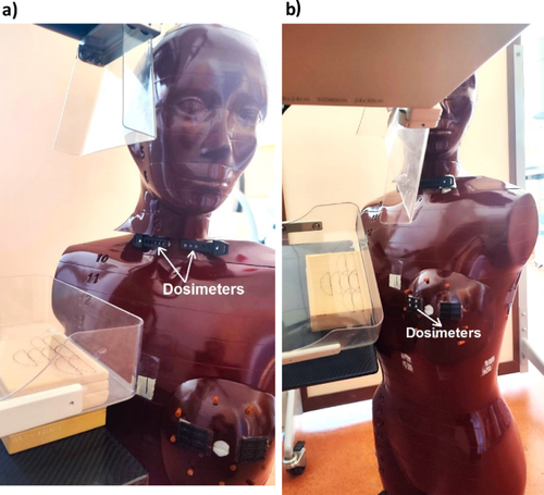
The organs of the thyroid and contralateral breast can be exposed to radiation because of scattered x-rays during mammography. It is important to determine the dose values of these quantities in terms of the risk of triggering a second cancer induction for both organs. In the present study, thyroid and contralateral breast surface doses were investigated with LiF:Mg,Ti (TLD-100) dosimeters for three types of BR-12 phantoms with glandularity/fat tissue ratios (70%/30%, 50%/50% and 30%/70%) over four views as in the real patient situation in Mo/Mo and Mo/Rh anode/filter combinations in each phantom thickness as opposed to a single glandular/fat tissue commonly used in the literature. Both thyroid and contralateral surface doses increased with increasing both phantom thickness and glandularity/fat ratio. The thyroid surface doses changed within 0.06–0.18 mGy and 0.05–0.14 mGy according to phantom thickness of 2 to 6 cm for the glandularity/adipose of 50%/50% in the Mo/Mo and Mo/Rh, respectively. On the other hand, the contralateral breast surface doses were within 0.35–1.39 mGy and 0.40–0.99 mGy for 50%/50% in the Mo/Mo and Mo/Rh, respectively. Based on a 70%/30% breast composition with a phantom thickness of 6 cm, the thyroid and contralateral breast surface doses increased by approximately 40% compared with 50%/50% for both Mo/Mo and Mo/Rh. These results showed that both thyroid and contralateral breast surface doses significantly depend on the glandularity/fat composition of the breast. Therefore, the outputs of this study may contribute to future studies aimed at reducing the doses received by organs during examination.
中文翻译:
乳房X线照相术中甲状腺和对侧乳房表面剂量变化:关于乳腺组织成分作用的模型研究
甲状腺和对侧乳房的器官可能会因乳房 X 光检查过程中 X 射线的散射而暴露在辐射下。根据触发两个器官第二次癌症诱导的风险来确定这些量的剂量值非常重要。在本研究中,使用 LiF:Mg,Ti (TLD-100) 剂量计对三种类型 BR-12 模型的腺体/脂肪组织比率(70%/30%、50%/50)研究了甲状腺和对侧乳房表面剂量。 %和30%/70%)超过四个视图,就像在每个体模厚度中的Mo/Mo和Mo/Rh阳极/过滤器组合中的真实患者情况一样,而不是文献中常用的单个腺体/脂肪组织。甲状腺和对侧表面剂量都随着体模厚度和腺体/脂肪比率的增加而增加。对于Mo/Mo和Mo/Rh中50%/50%的腺体/脂肪,甲状腺表面剂量根据2至6cm的体模厚度分别在0.06-0.18 mGy和0.05-0.14 mGy内变化。另一方面,Mo/Mo 和 Mo/Rh 中 50%/50% 的对侧乳房表面剂量分别在 0.35-1.39 mGy 和 0.40-0.99 mGy 范围内。基于体模厚度为 6 cm 的 70%/30% 乳房成分,甲状腺和对侧乳房表面剂量增加了约 40%,而 Mo/Mo 和 Mo/Rh 的剂量均为 50%/50%。这些结果表明,甲状腺和对侧乳房表面剂量显着取决于乳房的腺体/脂肪成分。因此,这项研究的结果可能有助于未来旨在减少检查过程中器官接受的剂量的研究。


 京公网安备 11010802027423号