Journal of Cardiovascular Translational Research ( IF 3.4 ) Pub Date : 2024-04-16 , DOI: 10.1007/s12265-024-10505-x Renee C. Brigham , Alexander R. Mattson , Paul A. Iaizzo
|
Epicardial interventions have forged new frontiers in cardiac ablation and device therapies. Healthy human hearts typically present with significant adipose tissue layers superficial to the ventricular myocardium and may hinder success or increase the complexities of epicardial interventions. We quantitatively evaluated the distribution of epicardial adipose tissue on the surface of human hearts and provided high-fidelity 3-dimensional reconstructions of these epicardial adipose tissue layers. The regional thickness of adipose tissues was analyzed at 51 anatomical reference points surrounding both ventricles and compared to specific patient demographics. Adipose deposits on the human hearts displayed characteristic patterns, with the thickest accumulations along the interventricular septa (anterior, 9.01 ± 0.50 mm; posterior, 6.78 ± 0.50 mm) and the right ventricular margin (7.44 ± 0.57 mm). We provide one of the most complete characterizations of human epicardial adipose location and relative layer thickness. These results are considered fundamental for an underlying anatomic understanding when performing procedures within the pericardial space.
Graphical Abstract
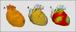
The relative thickness of epicardial adipose tissue was analyzed across 80 human hearts, with a subset displayed here as 3D reconstructions with thinner to thicker adipose regions indicated by a relative green-to-red color scale.
中文翻译:
人类心脏的心室心外膜脂肪分布:3 维重建和定量评估
心外膜干预开辟了心脏消融和设备治疗的新领域。健康的人类心脏通常在心室心肌表面存在大量脂肪组织层,可能会阻碍心外膜干预的成功或增加其复杂性。我们定量评估了人类心脏表面心外膜脂肪组织的分布,并提供了这些心外膜脂肪组织层的高保真度三维重建。在两个心室周围的 51 个解剖参考点分析脂肪组织的区域厚度,并与特定患者的人口统计数据进行比较。人类心脏上的脂肪沉积表现出特征性模式,沿室间隔(前部,9.01±0.50毫米;后部,6.78±0.50毫米)和右心室边缘(7.44±0.57毫米)的脂肪沉积最厚。我们提供人类心外膜脂肪位置和相对层厚度的最完整特征之一。这些结果被认为是在心包腔内进行手术时了解基本解剖学的基础。
图形概要
对 80 个人类心脏的心外膜脂肪组织的相对厚度进行了分析,其中一个子集在此显示为 3D 重建,其中由相对绿到红的色阶表示从薄到厚的脂肪区域。


 京公网安备 11010802027423号