当前位置:
X-MOL 学术
›
Acc. Chem. Res.
›
论文详情
Our official English website, www.x-mol.net, welcomes your feedback! (Note: you will need to create a separate account there.)
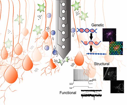
Structural, Functional, and Genetic Changes Surrounding Electrodes Implanted in the Brain
Accounts of Chemical Research ( IF 18.3 ) Pub Date : 2024-04-17 , DOI: 10.1021/acs.accounts.4c00057 Bhavna Gupta 1, 2 , Akash Saxena 2, 3 , Mason L. Perillo 2, 4 , Lauren C. Wade-Kleyn 2, 4 , Cort H. Thompson 2, 4 , Erin K. Purcell 1, 2, 3, 4
Accounts of Chemical Research ( IF 18.3 ) Pub Date : 2024-04-17 , DOI: 10.1021/acs.accounts.4c00057 Bhavna Gupta 1, 2 , Akash Saxena 2, 3 , Mason L. Perillo 2, 4 , Lauren C. Wade-Kleyn 2, 4 , Cort H. Thompson 2, 4 , Erin K. Purcell 1, 2, 3, 4
Affiliation
|
Implantable neurotechnology enables monitoring and stimulating of the brain signals responsible for performing cognitive, motor, and sensory tasks. Electrode arrays implanted in the brain are increasingly used in the clinic to treat a variety of sources of neurological diseases and injuries. However, the implantation of a foreign body typically initiates a tissue response characterized by physical disruption of vasculature and the neuropil as well as the initiation of inflammation and the induction of reactive glial states. Likewise, electrical stimulation can induce damage to the surrounding tissue depending on the intensity and waveform parameters of the applied stimulus. These phenomena, in turn, are likely influenced by the surface chemistry and characteristics of the materials employed, but further information is needed to effectively link the biological responses observed to specific aspects of device design. In order to inform improved design of implantable neurotechnology, we are investigating the basic science principles governing device–tissue integration. We are employing multiple techniques to characterize the structural, functional, and genetic changes that occur in the cells surrounding implanted electrodes. First, we have developed a new “device-in-slice” technique to capture chronically implanted electrodes within thick slices of live rat brain tissue for interrogation with single-cell electrophysiology and two-photon imaging techniques. Our data revealed several new observations of tissue remodeling surrounding devices: (a) there was significant disruption of dendritic arbors in neurons near implants, where losses were driven asymmetrically on the implant-facing side. (b) There was a significant loss of dendritic spine densities in neurons near implants, with a shift toward more immature (nonfunctional) morphologies. (c) There was a reduction in excitatory neurotransmission surrounding implants, as evidenced by a reduction in the frequency of excitatory postsynaptic currents (EPSCs). Lastly, (d) there were changes in the electrophysiological underpinnings of neuronal spiking regularity. In parallel, we initiated new studies to explore changes in gene expression surrounding devices through spatial transcriptomics, which we applied to both recording and stimulating arrays. We found that (a) device implantation is associated with the induction of hundreds of genes associated with neuroinflammation, glial reactivity, oligodendrocyte function, and cellular metabolism and (b) electrical stimulation induces gene expression associated with damage or plasticity in a manner dependent upon the intensity of the applied stimulus. We are currently developing computational analysis tools to distill biomarkers of device–tissue interactions from large transcriptomics data sets. These results improve the current understanding of the biological response to electrodes implanted in the brain while producing new biomarkers for benchmarking the effects of novel electrode designs on responses. As the next generation of neurotechnology is developed, it will be increasingly important to understand the influence of novel materials, surface chemistries, and implant architectures on device performance as well as the relationship with the induction of specific cellular signaling pathways.
中文翻译:

植入大脑的电极周围的结构、功能和遗传变化
植入式神经技术能够监测和刺激负责执行认知、运动和感觉任务的大脑信号。植入大脑的电极阵列越来越多地用于临床治疗各种来源的神经系统疾病和损伤。然而,异物的植入通常会引发组织反应,其特征是脉管系统和神经纤维的物理破坏以及炎症的引发和反应性神经胶质状态的诱导。同样,电刺激可能会对周围组织造成损害,具体取决于所施加刺激的强度和波形参数。反过来,这些现象可能受到所用材料的表面化学和特性的影响,但需要进一步的信息来有效地将观察到的生物反应与设备设计的特定方面联系起来。为了为植入式神经技术的改进设计提供信息,我们正在研究控制设备与组织整合的基本科学原理。我们正在采用多种技术来表征植入电极周围细胞中发生的结构、功能和遗传变化。首先,我们开发了一种新的“切片设备”技术,可以在活体大鼠脑组织的厚切片中捕获长期植入的电极,以便用单细胞电生理学和双光子成像技术进行询问。我们的数据揭示了有关装置周围组织重塑的几个新观察结果:(a)植入物附近的神经元中的树突状乔木受到显着破坏,其中损失在面向植入物的一侧不对称地驱动。 (b) 植入物附近的神经元中树突棘密度显着损失,并转向更不成熟(无功能)的形态。 (c) 植入物周围的兴奋性神经传递减少,兴奋性突触后电流 (EPSC) 频率的减少证明了这一点。最后,(d)神经元尖峰规律的电生理学基础发生了变化。与此同时,我们发起了新的研究,通过空间转录组学探索设备周围基因表达的变化,并将其应用于记录和刺激阵列。我们发现(a)装置植入与数百种与神经炎症、神经胶质反应性、少突胶质细胞功能和细胞代谢相关的基因的诱导有关,(b)电刺激以依赖于损伤或可塑性的方式诱导与损伤或可塑性相关的基因表达。所施加刺激的强度。我们目前正在开发计算分析工具,以从大型转录组数据集中提取设备与组织相互作用的生物标志物。这些结果提高了目前对植入大脑的电极的生物反应的理解,同时产生了新的生物标志物,用于对新型电极设计对反应的影响进行基准测试。随着下一代神经技术的发展,了解新型材料、表面化学和植入物结构对设备性能的影响以及与特定细胞信号通路诱导的关系将变得越来越重要。
更新日期:2024-04-18
中文翻译:
植入大脑的电极周围的结构、功能和遗传变化
植入式神经技术能够监测和刺激负责执行认知、运动和感觉任务的大脑信号。植入大脑的电极阵列越来越多地用于临床治疗各种来源的神经系统疾病和损伤。然而,异物的植入通常会引发组织反应,其特征是脉管系统和神经纤维的物理破坏以及炎症的引发和反应性神经胶质状态的诱导。同样,电刺激可能会对周围组织造成损害,具体取决于所施加刺激的强度和波形参数。反过来,这些现象可能受到所用材料的表面化学和特性的影响,但需要进一步的信息来有效地将观察到的生物反应与设备设计的特定方面联系起来。为了为植入式神经技术的改进设计提供信息,我们正在研究控制设备与组织整合的基本科学原理。我们正在采用多种技术来表征植入电极周围细胞中发生的结构、功能和遗传变化。首先,我们开发了一种新的“切片设备”技术,可以在活体大鼠脑组织的厚切片中捕获长期植入的电极,以便用单细胞电生理学和双光子成像技术进行询问。我们的数据揭示了有关装置周围组织重塑的几个新观察结果:(a)植入物附近的神经元中的树突状乔木受到显着破坏,其中损失在面向植入物的一侧不对称地驱动。 (b) 植入物附近的神经元中树突棘密度显着损失,并转向更不成熟(无功能)的形态。 (c) 植入物周围的兴奋性神经传递减少,兴奋性突触后电流 (EPSC) 频率的减少证明了这一点。最后,(d)神经元尖峰规律的电生理学基础发生了变化。与此同时,我们发起了新的研究,通过空间转录组学探索设备周围基因表达的变化,并将其应用于记录和刺激阵列。我们发现(a)装置植入与数百种与神经炎症、神经胶质反应性、少突胶质细胞功能和细胞代谢相关的基因的诱导有关,(b)电刺激以依赖于损伤或可塑性的方式诱导与损伤或可塑性相关的基因表达。所施加刺激的强度。我们目前正在开发计算分析工具,以从大型转录组数据集中提取设备与组织相互作用的生物标志物。这些结果提高了目前对植入大脑的电极的生物反应的理解,同时产生了新的生物标志物,用于对新型电极设计对反应的影响进行基准测试。随着下一代神经技术的发展,了解新型材料、表面化学和植入物结构对设备性能的影响以及与特定细胞信号通路诱导的关系将变得越来越重要。


 京公网安备 11010802027423号