Our official English website, www.x-mol.net, welcomes your feedback! (Note: you will need to create a separate account there.)
Revealing the MRI‐Contrast in Optically Cleared Brains
Advanced Science ( IF 15.1 ) Pub Date : 2024-04-22 , DOI: 10.1002/advs.202400316 Shimrit Oz 1 , Galit Saar 2 , Shunit Olszakier 1 , Ronit Heinrich 1 , Mykhail O. Kompanets 3 , Shai Berlin 1
Advanced Science ( IF 15.1 ) Pub Date : 2024-04-22 , DOI: 10.1002/advs.202400316 Shimrit Oz 1 , Galit Saar 2 , Shunit Olszakier 1 , Ronit Heinrich 1 , Mykhail O. Kompanets 3 , Shai Berlin 1
Affiliation
|
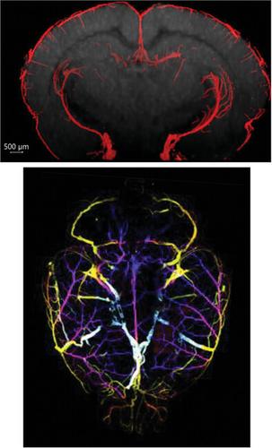
The current consensus holds that optically‐cleared specimens are unsuitable for Magnetic Resonance Imaging (MRI); exhibiting absence of contrast. Prior studies combined MRI with tissue‐clearing techniques relying on the latter's ability to eliminate lipids, thereby fostering the assumption that lipids constitute the primary source of ex vivo MRI‐contrast. Nevertheless, these findings contradict an extensive body of literature that underscores the contribution of other features to contrast. Furthermore, it remains unknown whether non‐delipidating clearing methods can produce MRI‐compatible specimens or whether MRI‐contrast can be re‐established. These limitations hinder the development of multimodal MRI‐light‐microscopy (LM) imaging approaches. This study assesses the relation between MRI‐contrast, and delipidation in optically‐cleared whole brains following different tissue‐clearing approaches. It is demonstrated that uDISCO and ECi‐brains are MRI‐compatible upon tissue rehydration, despite both methods’ substantial delipidating‐nature. It is also demonstrated that, whereas Scal e‐clearing preserves most lipids, Scal e‐cleared brain lack MRI‐contrast. Furthermore, MRI‐contrast is restored to lipid‐free CLARITY‐brains without introducing lipids. Our results thereby dissociate between the essentiality of lipids to MRI‐contrast. A tight association is found between tissue expansion, hyperhydration and loss of MRI‐contrast. These findings then enabled us to develop a multimodal MRI‐LM‐imaging approach, opening new avenues to bridge between the micro‐ and mesoscale for biomedical research and clinical applications.
中文翻译:

揭示光学透明大脑中的 MRI 对比
目前的共识认为光学透明的标本不适合磁共振成像(MRI);表现出缺乏对比。先前的研究将 MRI 与组织清除技术相结合,依赖于后者消除脂质的能力,从而促进了脂质构成离体 MRI 对比的主要来源的假设。然而,这些发现与大量强调其他特征对对比的贡献的文献相矛盾。此外,目前尚不清楚非脱脂透明方法是否可以产生与 MRI 兼容的标本,或者是否可以重新建立 MRI 对比。这些限制阻碍了多模态 MRI 光学显微镜 (LM) 成像方法的发展。本研究评估了 MRI 对比度与采用不同组织透明化方法的光学透明全脑脱脂之间的关系。事实证明,uDISCO 和 ECi-brain 在组织补液时与 MRI 兼容,尽管这两种方法都具有显着的脱脂性质。还证明,虽然 Sca我 电子清算保留了大部分脂质,Sca我 电子清除的大脑缺乏 MRI 对比。此外,在不引入脂质的情况下,MRI 对比可以恢复到无脂质的 CLARITY 大脑。因此,我们的结果区分了脂质与 MRI 对比的重要性。研究发现组织扩张、过度水合和 MRI 对比度丧失之间存在密切关联。这些发现使我们能够开发出一种多模态 MRI-LM 成像方法,为生物医学研究和临床应用的微观和介观尺度之间的桥梁开辟了新的途径。
更新日期:2024-04-22
中文翻译:
揭示光学透明大脑中的 MRI 对比
目前的共识认为光学透明的标本不适合磁共振成像(MRI);表现出缺乏对比。先前的研究将 MRI 与组织清除技术相结合,依赖于后者消除脂质的能力,从而促进了脂质构成离体 MRI 对比的主要来源的假设。然而,这些发现与大量强调其他特征对对比的贡献的文献相矛盾。此外,目前尚不清楚非脱脂透明方法是否可以产生与 MRI 兼容的标本,或者是否可以重新建立 MRI 对比。这些限制阻碍了多模态 MRI 光学显微镜 (LM) 成像方法的发展。本研究评估了 MRI 对比度与采用不同组织透明化方法的光学透明全脑脱脂之间的关系。事实证明,uDISCO 和 ECi-brain 在组织补液时与 MRI 兼容,尽管这两种方法都具有显着的脱脂性质。还证明,虽然 Sca


 京公网安备 11010802027423号