Cellular Oncology ( IF 6.6 ) Pub Date : 2024-04-23 , DOI: 10.1007/s13402-024-00949-3 Yujing Huang , Dongyan Cao , Manxue Zhang , Yue Yang , Gengming Niu , Lina Tang , Zan Shen , Zhichang Zhang , Yueqing Bai , Daliu Min , Aina He
|
Purpose
The overall survival rate for metastatic osteosarcoma hovers around 20%. Responses to second-line chemotherapy, targeted therapies, and immunotherapies have demonstrated limited efficacy in metastatic osteosarcoma. Our objective is to validate differentially expressed genes and signaling pathways between non-metastatic and metastatic osteosarcoma, employing single-cell RNA sequencing (scRNA-seq) and additional functional investigations. We aim to enhance comprehension of metastatic mechanisms and potentially unveil a therapeutic target.
Methods
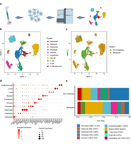
scRNA-seq was performed on two primary osteosarcoma lesions (1 non-metastatic and 1 metastatic). Seurat package facilitated dimensionality reduction and cluster identification. Copy number variation (CNV) was predicted using InferCNV. CellChat characterized ligand-receptor-based intercellular communication networks. Differentially expressed genes underwent GO function enrichment analysis and GSEA. Validation was achieved through the GSE152048 dataset, which identified PDGFD-PDGFRB as a common ligand-receptor pair with significant contribution. Immunohistochemistry assessed PDGFD and PDGFRB expression, while multicolor immunofluorescence and flow cytometry provided insight into spatial relationships and the tumor immune microenvironment. Kaplan-Meier survival analysis compared metastasis-free survival and overall survival between high and low levels of PDGFD and PDGFRB. Manipulation of PDGFD expression in primary osteosarcoma cells examined invasion abilities and related markers.
Results
Ten clusters encompassing osteoblasts, osteoclasts, osteocytes, fibroblasts, pericytes, endothelial cells, myeloid cells, T cells, B cells, and proliferating cells were identified. Osteoblasts, osteoclasts, and osteocytes exhibited heightened CNV levels. Ligand-receptor-based communication networks exposed significant fibroblast crosstalk with other cell types, and the PDGF signaling pathway was activated in non-metastatic osteosarcoma primary lesion. These results were corroborated by the GSE152048 dataset, confirming the prominence of PDGFD-PDGFRB as a common ligand-receptor pair. Immunohistochemistry demonstrated considerably greater PDGFD expression in non-metastatic osteosarcoma tissues and organoids, correlating with extended metastasis-free and overall survival. PDGFRB expression showed no significant variation between non-metastatic and metastatic osteosarcoma, nor strong correlations with survival times. Multicolor immunofluorescence suggested co-localization of PDGFD with PDGFRB. Flow cytometry unveiled a highly immunosuppressive microenvironment in metastatic osteosarcoma. Manipulating PDGFD expression demonstrated altered invasive abilities and marker expressions in primary osteosarcoma cells from both non-metastatic and metastatic lesions.
Conclusions
scRNA-seq illuminated the activation of the PDGF signaling pathway in primary lesion of non-metastatic osteosarcoma. PDGFD displayed an inhibitory effect on osteosarcoma metastasis, likely through the suppression of the EMT signaling pathway.
中文翻译:
通过单细胞测序分析探讨PDGFD对骨肉瘤转移的影响
目的
转移性骨肉瘤的总生存率徘徊在 20% 左右。二线化疗、靶向治疗和免疫疗法对转移性骨肉瘤的疗效有限。我们的目标是利用单细胞 RNA 测序 (scRNA-seq) 和其他功能研究来验证非转移性和转移性骨肉瘤之间的差异表达基因和信号通路。我们的目标是增强对转移机制的理解并可能揭示治疗靶点。
方法
对两个原发性骨肉瘤病变(1 个非转移性和 1 个转移性)进行了 scRNA-seq。 Seurat 包促进了降维和聚类识别。使用 InferCNV 预测拷贝数变异 (CNV)。 CellChat 描述了基于配体受体的细胞间通讯网络。对差异表达基因进行GO功能富集分析和GSEA。通过 GSE152048 数据集进行了验证,该数据集将 PDGFD-PDGFRB 确定为具有重大贡献的常见配体-受体对。免疫组织化学评估了 PDGFD 和 PDGFRB 的表达,而多色免疫荧光和流式细胞术则提供了对空间关系和肿瘤免疫微环境的深入了解。 Kaplan-Meier 生存分析比较了高水平和低水平 PDGFD 和 PDGFRB 之间的无转移生存和总生存。原代骨肉瘤细胞中 PDGFD 表达的操作检查了侵袭能力和相关标记。
结果
鉴定出包括成骨细胞、破骨细胞、骨细胞、成纤维细胞、周细胞、内皮细胞、骨髓细胞、T细胞、B细胞和增殖细胞的十个簇。成骨细胞、破骨细胞和骨细胞表现出升高的 CNV 水平。基于配体受体的通讯网络暴露出与其他细胞类型显着的成纤维细胞串扰,并且PDGF信号通路在非转移性骨肉瘤原发性病变中被激活。这些结果得到了 GSE152048 数据集的证实,证实了 PDGFD-PDGFRB 作为常见配体-受体对的重要性。免疫组织化学显示,非转移性骨肉瘤组织和类器官中 PDGFD 表达显着升高,与延长的无转移生存期和总生存期相关。 PDGFRB 表达在非转移性和转移性骨肉瘤之间没有显着差异,也与生存时间没有很强的相关性。多色免疫荧光表明 PDGFD 与 PDGFRB 共定位。流式细胞术揭示了转移性骨肉瘤中高度免疫抑制的微环境。操纵PDGFD表达证明了来自非转移性和转移性病变的原发性骨肉瘤细胞的侵袭能力和标记物表达的改变。
结论
scRNA-seq 阐明了非转移性骨肉瘤原发灶中 PDGF 信号通路的激活。 PDGFD 对骨肉瘤转移具有抑制作用,可能是通过抑制 EMT 信号通路来实现的。


 京公网安备 11010802027423号