当前位置:
X-MOL 学术
›
J. Autoimmun.
›
论文详情
Our official English website, www.x-mol.net, welcomes your feedback! (Note: you will need to create a separate account there.)
Impaired IL-6-induced JAK-STAT signaling in CD4+ T cells associates with longer treatment duration in giant cell arteritis
Journal of Autoimmunity ( IF 12.8 ) Pub Date : 2024-04-22 , DOI: 10.1016/j.jaut.2024.103215 Idil Esen , Maria Sandovici , Peter Heeringa , Annemieke M.H. Boots , Elisabeth Brouwer , Yannick van Sleen , Wayel Abdulahad
Journal of Autoimmunity ( IF 12.8 ) Pub Date : 2024-04-22 , DOI: 10.1016/j.jaut.2024.103215 Idil Esen , Maria Sandovici , Peter Heeringa , Annemieke M.H. Boots , Elisabeth Brouwer , Yannick van Sleen , Wayel Abdulahad
|
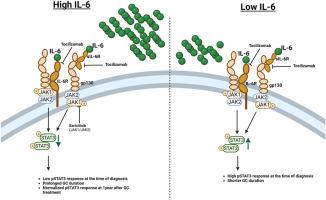
The IL-12-IFNγ-Th1 and the IL-6-IL-23-Th17 axes are considered the dominant pathogenic pathways in Giant Cell Arteritis (GCA). Both pathways signal via activation of the downstream JAK/STAT proteins. We hypothesized that phosphorylated STAT (pSTAT) signatures in circulating immune cells may aid to stratify GCA-patients for personalized treatment. To investigate pSTAT expression, PBMCs from treatment-naive GCA-patients (n = 18), infection controls (INF, n = 11) and age-matched healthy controls (HC, n = 15) were stimulated in vitro with IL-6, IL-2, IL-10, IFN-γ, M-CSF or GM-CSF, and stained with CD3, CD4, CD19, CD45RO, pSTAT1, pSTAT3, pSTAT5 antibodies, and analyzed by flow cytometry. Serum IL-6, sIL-6-receptor and gp130 were measured by Luminex. The change in percentages of pSTAT3+CD4+T-cells was evaluated at diagnosis and at 3 months and 1-year of follow-up. Kaplan-Meier analyses was used to asses prognostic accuracy. Analysis of IL-6 stimulated immune cell subsets revealed a significant decrease in percentages of pSTAT3+CD4+T-cells of GCA-patients and INF-controls compared to HCs. Following patient stratification according to high (median>1.5 pg/mL) and low (median<1.5 pg/mL) IL-6 levels, we observed a reduction in the pSTAT3 response in GCA-patients with high serum IL-6. Percentages of pSTAT3+CD4+T-cells in patients with high serum IL-6 levels at diagnosis normalized after glucocorticoid (GC) treatment. Importantly, we found that patients with low percentages of pSTAT3+CD4+T-cells at baseline require longer GC-treatment. Overall, in GCA, the percentages of in vitro IL-6-induced pSTAT3+CD4+T-cells likely reflect prior in vivo exposure to high IL-6 and may serve as a prognostic marker for GC-treatment duration and may assist improving personalized treatment options in the future.
中文翻译:

CD4+ T 细胞中 IL-6 诱导的 JAK-STAT 信号传导受损与巨细胞动脉炎治疗持续时间延长相关
IL-12-IFNγ-Th1 和 IL-6-IL-23-Th17 轴被认为是巨细胞动脉炎 (GCA) 的主要致病途径。两条途径均通过下游 JAK/STAT 蛋白的激活发出信号。我们假设循环免疫细胞中的磷酸化 STAT (pSTAT) 特征可能有助于对 GCA 患者进行分层以进行个性化治疗。为了研究 pSTAT 表达,使用 IL-6 在体外刺激来自未接受治疗的 GCA 患者 (n = 18)、感染对照 (INF,n = 11) 和年龄匹配的健康对照 (HC,n = 15) 的 PBMC, IL-2、IL-10、IFN-γ、M-CSF 或 GM-CSF,并用 CD3、CD4、CD19、CD45RO、pSTAT1、pSTAT3、pSTAT5 抗体染色,并通过流式细胞术进行分析。通过Luminex测量血清IL-6、sIL-6受体和gp130。在诊断时以及 3 个月和 1 年随访时评估 pSTAT3+CD4+T 细胞百分比的变化。 Kaplan-Meier 分析用于评估预后准确性。对 IL-6 刺激的免疫细胞亚群的分析显示,与 HC 相比,GCA 患者和 INF 对照的 pSTAT3+CD4+T 细胞百分比显着下降。根据高(中位数>1.5 pg/mL)和低(中位数<1.5 pg/mL)IL-6水平对患者进行分层后,我们观察到血清IL-6高的GCA患者的pSTAT3反应降低。诊断时血清 IL-6 水平高的患者中 pSTAT3+CD4+T 细胞的百分比在糖皮质激素 (GC) 治疗后恢复正常。重要的是,我们发现基线时 pSTAT3+CD4+T 细胞百分比较低的患者需要更长的 GC 治疗。总体而言,在 GCA 中,体外 IL-6 诱导的 pSTAT3+CD4+T 细胞的百分比可能反映了先前体内暴露于高 IL-6 的情况,并且可以作为 GC 治疗持续时间的预后标志物,并可能有助于改善个性化治疗未来的治疗选择。
更新日期:2024-04-22
中文翻译:
CD4+ T 细胞中 IL-6 诱导的 JAK-STAT 信号传导受损与巨细胞动脉炎治疗持续时间延长相关
IL-12-IFNγ-Th1 和 IL-6-IL-23-Th17 轴被认为是巨细胞动脉炎 (GCA) 的主要致病途径。两条途径均通过下游 JAK/STAT 蛋白的激活发出信号。我们假设循环免疫细胞中的磷酸化 STAT (pSTAT) 特征可能有助于对 GCA 患者进行分层以进行个性化治疗。为了研究 pSTAT 表达,使用 IL-6 在体外刺激来自未接受治疗的 GCA 患者 (n = 18)、感染对照 (INF,n = 11) 和年龄匹配的健康对照 (HC,n = 15) 的 PBMC, IL-2、IL-10、IFN-γ、M-CSF 或 GM-CSF,并用 CD3、CD4、CD19、CD45RO、pSTAT1、pSTAT3、pSTAT5 抗体染色,并通过流式细胞术进行分析。通过Luminex测量血清IL-6、sIL-6受体和gp130。在诊断时以及 3 个月和 1 年随访时评估 pSTAT3+CD4+T 细胞百分比的变化。 Kaplan-Meier 分析用于评估预后准确性。对 IL-6 刺激的免疫细胞亚群的分析显示,与 HC 相比,GCA 患者和 INF 对照的 pSTAT3+CD4+T 细胞百分比显着下降。根据高(中位数>1.5 pg/mL)和低(中位数<1.5 pg/mL)IL-6水平对患者进行分层后,我们观察到血清IL-6高的GCA患者的pSTAT3反应降低。诊断时血清 IL-6 水平高的患者中 pSTAT3+CD4+T 细胞的百分比在糖皮质激素 (GC) 治疗后恢复正常。重要的是,我们发现基线时 pSTAT3+CD4+T 细胞百分比较低的患者需要更长的 GC 治疗。总体而言,在 GCA 中,体外 IL-6 诱导的 pSTAT3+CD4+T 细胞的百分比可能反映了先前体内暴露于高 IL-6 的情况,并且可以作为 GC 治疗持续时间的预后标志物,并可能有助于改善个性化治疗未来的治疗选择。


 京公网安备 11010802027423号