Immunofluorescence signal intensity measurements as a semi-quantitative tool to assess sarcoglycan complex expression in muscle biopsy

Submitted: 13 April 2022
Accepted: 20 June 2022
Published: 1 September 2022
Abstract Views: 615
PDF: 474
HTML: 17
Publisher's note
All claims expressed in this article are solely those of the authors and do not necessarily represent those of their affiliated organizations, or those of the publisher, the editors and the reviewers. Any product that may be evaluated in this article or claim that may be made by its manufacturer is not guaranteed or endorsed by the publisher.

Authors

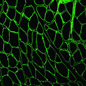
Sarcoglycanopathies are highly heterogeneous in terms of disease progression, muscular weakness, loss of ambulation and cardiac/respiratory involvement. Their clinical severity usually correlates with the residual protein amount, which makes protein quantification extremely relevant. Sarcoglycanopathy diagnosis is genetic, but skeletal muscle analysis - by both immunohistochemistry and Western blot (WB) - is still mandatory to establish the correct diagnostic process. Unfortunately, however, WB analysis cannot be performed if the bioptic specimen is scarce. This study provides a sensitive tool for semi-quantification of residual amount of sarcoglycans in patients affected by sarcoglycanopathies, based on immunofluorescence staining on skeletal muscle sections, image acquisition and software elaboration. We applied this method to eleven sarcoglycanopathies, seven Becker muscular dystrophies and four age-matched controls. Fluorescence data analysed in patients and compared to age-matched controls showed a significant reduction of the mutated sarcoglycan expression and a variable reduction of the other sarcoglycans. Fluorescence normalized data analysed in relation to the age of onset of the disease, showed a negative correlation of α-sarcoglycan fluorescent signal versus fibrosis in patients with an early age of onset and a negative correlation between δ-sarcoglycan signal and fibrosis in both intermediate and late age of onset groups. The availability of a method that allows objective quantification of the sarcolemmal proteins, faster and less consuming than WB analysis and able to detect low residual sarcoglycan expression with great sensitivity, proves useful to better define both patient prognosis and expected disease evolution. The proposed method could be employed also to monitor the efficacy of therapeutic interventions and during clinical trials.

Dimensions

Altmetric

PlumX Metrics

Downloads

Download data is not yet available.

Citations

Roberds SL, Anderson RD, Ibraghimov-Beskrovnaya O, Campbell, K.P. Primary structure and muscle-specific expression of the 50-kDa dystrophin-associated glycoprotein (adhalin). J Biol Chem 1993;268:23739-42. DOI: https://doi.org/10.1016/S0021-9258(20)80440-2
Lim LE, Duclos F, Broux O, Bourg N, Sunada Y, Allamand V, et al. Beta-sarcoglycan: characterization and role in limb-girdle muscular dystrophy linked to 4q12. Nat Genet 1995;11:57-65. DOI: https://doi.org/10.1038/ng1195-257
Noguchi S, McNally EM, Ben Othmane K, Hagiwara Y, Mizuno Y, Yoshida M, et al. Mutations in the dystrophin-associated protein gamma-sarcoglycan in chromosome 13 muscular dystrophy. Science 1995;270:819-22. DOI: https://doi.org/10.1126/science.270.5237.819
Nigro V, Piluso G, Belsito A, Politano L, Puca AA, Papparella S, et al. Identification of a novel sarcoglycan gene at 5q33 encoding a sarcolemmal 35 kDa glycoprotein. Hum Mol Genet 1996;5:1179-86. DOI: https://doi.org/10.1093/hmg/5.8.1179
Holt KH, Campbell KP. Assembly of the sarcoglycan complex. Insights for muscular dystrophy. J Biol Chem 1998;273:34667-70. DOI: https://doi.org/10.1074/jbc.273.52.34667
Hack AA, Lam MY, Cordier L, Shoturma DI, Ly CT, Hadhazy MA, et al. Differential requirement for individual sarcoglycans and dystrophin in the assembly and function of the dystrophin-glycoprotein complex. J Cell Sci 2000;113:2535-44. DOI: https://doi.org/10.1242/jcs.113.14.2535
Guimarães-Costa R, Fernández-Eulate G, Wahbi K, Leturcq F, Malfatti E, Behin A, et al. Clinical correlations and long-term follow-up in 100 patients with sarcoglycanopathies. Eur J Neurol 2021;28:660-9. DOI: https://doi.org/10.1111/ene.14592
Vainzof M, Passos-Bueno MR, Canovas M, Moreira ES, Pavanello RC, Marie SK, et al. The sarcoglycan complex in the six autosomal recessive limb-girdle muscular dystrophies. Hum Mol Genet 1996;5:1963-9. DOI: https://doi.org/10.1093/hmg/5.12.1963
Mendell JR, Chicoine LG, Al-Zaidy SA, Sahenk Z, Lehman K, Lowes L, et al. Gene delivery for Limb-Girdle muscular dystrophy type 2D by isolated limb infusion. Hum Gene Ther 2019;30:794-801. DOI: https://doi.org/10.1089/hum.2019.006
Herson S, Hentati F, Rigolet A, Behin A, Romero NB, Leturcq F, et al. A phase I trial of adeno-associated virus serotype 1-gamma-sarcoglycan gene therapy for limb girdle muscular dystrophy type 2C. Brain 2012;135:483-92. DOI: https://doi.org/10.1093/brain/awr342
Peverelli L, Testolin S, Villa L, D'Amico A, Petrini S, Favero C, et al. M. Histologic muscular history in steroid-treated and untreated patients with Duchenne dystrophy. Neurology 2015;85:1886-93. DOI: https://doi.org/10.1212/WNL.0000000000002147
Bland JM, Altman DGJ. Agreement between methods of measurement with multiple observations per individual. Biopharm Stat 2007;17:571-82. DOI: https://doi.org/10.1080/10543400701329422
Sardone V, Ellis M, Torelli S, Feng L, Chambers D, Eastwood D, et al. A novel high-throughput immunofluorescence analysis method for quantifying dystrophin intensity in entire transverse sections of Duchenne muscular dystrophy muscle biopsy samples. PLoS One 2018;13:e0194540. DOI: https://doi.org/10.1371/journal.pone.0194540
Vainzof M, Souza LS, Gurgel-Giannetti J, Zatz M. Sarcoglycanopathies: an update. Neuromuscul Disord 2021;1021-7. DOI: https://doi.org/10.1016/j.nmd.2021.07.014
Scaglioni D, Ellis M, Catapano F, Torelli S, Chambers D, Feng L, et al. A high-throughput digital script for multiplexed immunofluorescent analysis and quantification of sarcolemmal and sarcomeric proteins in muscular dystrophies. Acta Neuropathol Commun 2020;8:53. DOI: https://doi.org/10.1186/s40478-020-00918-5
Beekman C, Sipkens JA, Testerink J, Giannakopoulos S, Kreuger D, van Deutekom JC, et al. A sensitive, reproducible and objective immunofluorescence analysis method of dystrophin in individual fibers in samples from patients with Duchenne muscular dystrophy. PLoS One 2014;9:e107494. DOI: https://doi.org/10.1371/journal.pone.0107494
Arechavala-Gomeza V, Kinali M, Feng L, Brown SC, Sewry C, Morgan JE, Muntoni F. Immunohistological intensity measurements as a tool to assess sarcolemma-associated protein expression. Neuropathol Appl Neurobiol 2010;36:265-74. DOI: https://doi.org/10.1111/j.1365-2990.2009.01056.x
Aeffner F, Faelan C, Moore SA, Moody A, Black JC, Charleston JS, et al. Validation of a muscle-specific tissue image analysis tool for quantitative assessment of dystrophin staining in frozen muscle biopsies. Arch Pathol Lab Med 2019;143:197-205. DOI: https://doi.org/10.5858/arpa.2017-0536-OA
Guglieri M, Magri F, D'Angelo MG, Prelle A, Morandi L, Rodolico C, et al. Clinical, molecular, and protein correlations in a large sample of genetically diagnosed Italian limb girdle muscular dystrophy patients. Hum Mutat 2008;29:258-66. DOI: https://doi.org/10.1002/humu.20642
Alonso-Pérez J, González-Quereda L, Bello L, Guglieri M, Straub V, Gallano P, et al. New genotype-phenotype correlations in a large European cohort of patients with sarcoglycanopathy. Brain 2020;143:2696-708. DOI: https://doi.org/10.1093/brain/awaa228
Giacomo Pietro Comi, Neuromuscular and Rare Disease Unit, Fondazione IRCCS Ca’ Granda Ospedale Maggiore Policlinico, Milan

Neurology Unit, Fondazione IRCCS Ca’ Granda Ospedale Maggiore Policlinico, Milan; Dino Ferrari Centre, Department of Pathophysiology and Transplantation (DEPT), University of Milan

How to Cite

Zanotti, S., Magri, F., Poggetti, F., Ripolone, M., Velardo, D., Fortunato, F., Ciscato, P., Moggio, M., Corti, S., Comi, G. P., & Sciacco, M. (2022). Immunofluorescence signal intensity measurements as a semi-quantitative tool to assess sarcoglycan complex expression in muscle biopsy. European Journal of Histochemistry, 66(3). https://doi.org/10.4081/ejh.2022.3418