

Abstract
The inherent biological hazards associated with ionizing radiation necessitate the implementation of effective shielding measures, particularly in medical applications. Interventional radiology, in particular, poses a unique challenge as it often exposes medical personnel to prolonged periods of high x-ray doses. Historically, lead and lead-based compounds have been the primary materials employed for shielding against photons. However, the drawbacks of lead, including its substantial weight causing personnel's inflexibility and its toxicity, have raised concerns regarding its long-term impact on both human health and the environment. Barium tantalate has emerged as a promising alternative, due to its unique attenuation properties against low-energy x-rays, specifically targeting the weak absorption area of lead. In the present study, we employ the Geant4 Monte Carlo simulation tool to investigate various formulations of barium tantalate doped with rare earth elements. The aim is to identify the optimal composition for shielding x-rays in the context of interventional radiology. To achieve this, we employ a reference x-ray spectrum typical of interventional radiology procedures, with energies extending up to 90 keV, within a carefully designed simulation setup. Our primary performance indicator is the reduction in air kerma transmission. Furthermore, we assess the absorbed doses to critical organs at risk within a standard human body phantom protected by the shield. Our results demonstrate that specific concentrations of the examined rare earth impurities can enhance the shielding performance of barium tantalate. To mitigate x-ray exposure in interventional radiology, our analysis reveals that the most effective shielding performance is achieved when using barium tantalate compositions containing 15% Erbium or 10% Samarium by weight. These findings suggest the possibility of developing lead-free shielding solutions or apron for interventional radiology personnel, offering a remarkable reduction in weight (exceeding 30%) while maintaining shielding performance at levels comparable to traditional lead-based materials.
Export citation and abstract BibTeX RIS

Original content from this work may be used under the terms of the Creative Commons Attribution 4.0 licence. Any further distribution of this work must maintain attribution to the author(s) and the title of the work, journal citation and DOI.
1. Introduction
With the expanding use of ionizing radiation in diagnostic imaging and radiation therapy, the importance of radiation protection has grown significantly to ensure the safety of both radiation workers and the general public. A fundamental principle of radiation protection involves the utilization of materials with exceptional radiation absorption and attenuation capabilities, commonly referred to as radiation shields. Traditionally, high atomic number metals like lead, tungsten, and bismuth, as well as their respective oxides, have been employed as shields against high-energy photons, including x-rays and gamma rays. Among these materials, lead and lead oxides have been the most prevalent choices for shielding against photons (He et al 2012, Jia et al 2016). However, concerns regarding the environmental toxicity of lead, its detrimental effects on human health, its substantial weight, and the associated high costs of waste disposal have raised global awareness about the challenges posed by its continued use (Sharma and Dubey 2005, Tchounwou et al 2012, Shik and Gholamzadeh 2018). Consequently, in recent years, extensive research efforts have been dedicated to the discovery of lead-free shielding compositions capable of effectively protecting against x-rays and gamma rays.
Interventional radiology, a pioneer in delivering image-guided diagnosis and treatment, combines advanced imaging techniques such as ultrasound and x-rays. Procedures involving x-rays, conducted through the use of fluoroscopy or CT scan equipment with energies ranging from 10–90 keV (Sabharwal et al 2007), typically require longer irradiation times compared to other radiological procedures. Additionally, interventional radiologists and staff often find themselves in closer proximity to the x-ray source compared to conventional x-ray imaging (Nowak et al 2020). Consequently, apart from the potential for patients to receive high doses of radiation (Ohuchi et al 2007), radiation shielding assumes paramount importance in interventional radiology procedures. Alarming reports have highlighted instances where up to 50% of radiology staff were exposed to doses exceeding the annual limit of 20 mSv, with considerably higher doses measured on protective aprons (Sánchez et al 2012). Furthermore, research has underscored the adverse health effects experienced by radiation workers who consistently wear lead aprons, including issues such as lower back pain, osteoarthritis, and increased pressure on the lumbar vertebrae (Moore et al 1992, Klein et al 2010). Given the extended duration of these procedures, there has been a growing impetus in research to explore alternative shielding materials that offer more favourable attributes, especially in terms of reduced toxicity, enhanced personnel's flexibility, and decreased weight (McCaffrey et al 2012, Kosaka et al 2019, Rajagopal and Anand 2022).
Although heavy metals are widely used for absorption and attenuation of radiation, in a range of low photon energies, they show a low absorption against X and gamma rays. This span or so-called feeble absorbing region for instance in case of lead is located in the range of 40–88 keV while lead shows a high absorption in the energy range 13–40 keV and beyond 80 keV (Jayakumar et al 2017, Wang et al 2020). Consequently, for x-ray spectra characterized by mean photon energies situated within this feeble absorbing region, the possibility arises to identify materials that can offer superior attenuation properties compared to pure lead (McCaffrey et al 2012). Take, for instance, bismuth, a non-toxic material with an atomic number of 83 but a lower mass density compared to lead. In the form of polymer-bismuth, it has been demonstrated as a viable alternative to lead, providing comparable photon attenuation characteristics, particularly in the low-energy region where photoelectric absorption predominates (Plionis et al 2009). Moreover, Chanthima et al (2012), in their investigation into the shielding properties of silicate glasses containing oxides of bismuth, barium, and lead, reported that the mass attenuation coefficient of the glass containing barium oxide surpasses those containing bismuth and lead within the energy range of 1–80 keV.
Composites are known with remarkable characteristics of low manufacturing costs and low weight. Due to its high atomic number and mass density (Z = 73, ρ = 16.69 g cm−3), tantalum stands out as a promising candidate for shielding against photons. In recent years, numerous tantalum-based compositions have emerged for radiation shielding applications (Yang et al 2015, Jia et al 2016). Among these compositions, barium tantalate (Ba5Ta4O15) has gained attention as a non-toxic material with excellent thermal and chemical stability, coupled with respectable radiation attenuation properties when compared to lead (Xu et al 2006, Jia et al 2016). Tantalum's unique characteristics come into focus when considering its x-ray absorption edges, with the L-edge at 11.68 keV and the K-edge at 67.42 keV. The feeble absorbing area falls between these energy values while tantalum shows a high absorption ability right above 67.42 keV. Furthermore, rare earth metals, ranging from lanthanum to lutetium, exhibit K-edges in the energy range of 38.9 to 63.3 keV (Bearden and Burr 1967). Incorporating these rare earth elements into the composition holds the potential to enhance the photon absorption capability of barium tantalate at energies below 67.4 keV. Consequently, it is anticipated that barium tantalate containing rare earth elements will demonstrate improved absorption properties in the lower-energy x-ray range applied in interventional radiology procedures when compared to uncontaminated material.
The Monte Carlo simulation is a well-established tool renowned for delivering reliable results in dosimetry and radiation shielding research, spanning both macroscopic and microscopic scales. It particularly proves invaluable when experimental approaches are either cost-prohibitive or unfeasible (Bozkurt and Bor 2006, Moradi et al 2019). Before this study, there has been a notable gap in obtaining detailed information regarding the impact of incorporating rare earth elements on the photon attenuation properties of barium tantalate composites. Consequently, this study aims to conduct a theoretical exploration of the x-ray shielding performance of Ba5Ta4O15 when augmented with varying concentrations of rare earth elements. The primary objective is to enhance the protection of interventional radiology personnel. To fulfil this objective, we employ Monte Carlo simulations utilizing the Geant4 code, thereby determining the optimal shield composition. We report here for the first time the use of barium tantalate containing various rare earth elements in order to optimize shielding performance tailored specifically for protection against x-rays encountered in interventional radiology. Additionally, we assess the absorbed doses to critical organs within a human phantom positioned under a protective apron, drawing comparisons with conventional lead shielding.
2. Material and methods
2.1. Geant4 Monte Carlo simulations
Geant4 stands out as a versatile Monte Carlo simulation tool freely accessible for various applications in radiation physics, medical sciences, nuclear physics, and optics. The most recent examples of Geant4 utilization in these domains can be found in the following publications: Broder et al 2022, Mohammadi et al 2022, Khodaei et al 2023, Taheri et al 2023. To assess the shielding efficacy of barium tantalate for interventional radiology applications, we employed the x-ray spectrum originally documented by Nowak et al in 2020 as a reference. In their study, x-ray energy spectra resulting from scattering within the patient's body and surrounding objects during an image-guided interventional procedure using an x-ray imaging system were meticulously measured. Measurements were conducted at various distances from the patient's examination table and at four distinct heights (170, 135, 96, and 53 cm, corresponding to eye, chest, belt, and knee level, respectively). These measurements were carried out using the Timepix3 chip which is a hybrid pixel detector developed at CERN having a silicon sensor with 500 μm thickness and separate readout for each pixel sensor. The highest radiation exposure was recorded at the radiologist's position, located 50 cm away from the x-ray tube at chest level (135 cm height). In our simulations, we digitized the photon energy spectrum corresponding to this specific point using the WebPlotDigitizer (accessible online at https://apps.automeris.io/wpd/), and this digitized spectrum served as our reference x-ray spectrum, illustrated in figure 1.
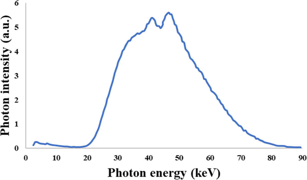
Figure 1. Photon energy spectrum measured at 50 cm distance from the x-ray tube in interventional radiology, sourced from the study by Nowak et al (2020).
Download figure:
Standard image High-resolution imageWe defined an x-ray source in the form of a flat, 4 cm diameter disk emitting a parallel photon beam with the spectrum shown in figure 1. This source was created using the G4GeneralParticleSource (GPS) tool and positioned at a 10 cm distance from the radiation shield. The shield, a flat plane measuring 20×20 cm2, had variable thicknesses ranging from 0.25 to 1 mm. Behind the shield, we placed a cylindrical detector with a 2 cm diameter and 2 cm height, situated 2 cm away from the shield surface. In each scenario, we calculated the photon spectrum recorded by the detector. A Geant4 visualization of the simulation geometry is demonstrated in figure 2.
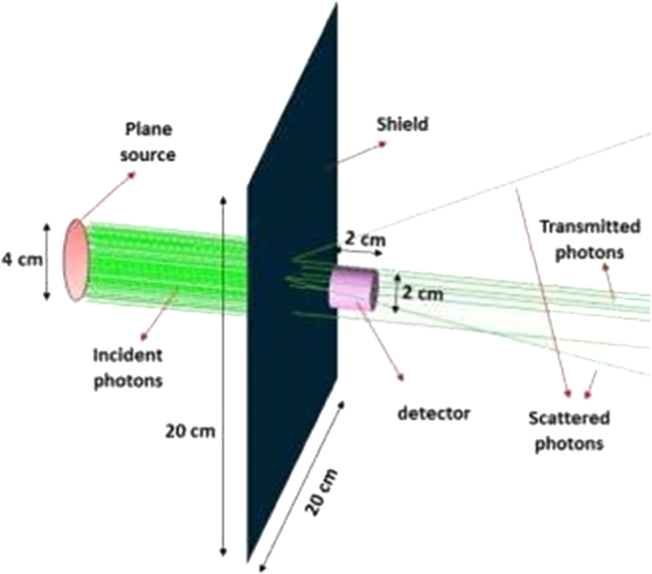
Figure 2. Geant4 visualization of the simulation geometry for 100 incident photons and 0.5 mm thickness shield.
Download figure:
Standard image High-resolution imageIn the simulations, we used Ba5Ta4O15 as the foundational material for the shield, which possesses a density of 7.69 g/cm3. This base material was combined with different rare earth impurities, including 1, 2, 5, 10, 12, and 15%. We initially opted for rare earth elements that are either non-radioactive or possess naturally occurring stable isotopes that are readily available in the market, either in metallic or oxide forms. The selection was limited to elements with purchase prices below 30 USD per kg, and these criteria included elements like 57La, 58Ce, 62Sm, 64Gd and 68Er. Subsequently, elements with higher procurement costs were introduced into the simulation analysis solely for the purpose of comparison. A list of these elements, along with their respective calculated densities, is provided in table 1. The characteristics of these shields will be contrasted with those of shields made of pure lead and unalloyed barium tantalate. It's worth noting that the varying fractions of impurities (as outlined in table 1) may lead to either a decrease or increase in density, which can result in either negative or positive effects on shielding performance. Additionally, due to the distinct feeble absorption characteristics of each element, the overall impact on shielding performance may be either favorable or unfavorable. The simulation results are anticipated to offer insights into the cumulative effect.
Table 1. Densities used in the simulations for various concentration of impurities within the Ba5Ta4O15 composition.
| Impurity | 57La | 58Ce | 59Pr | 60Nd | 62Sm | 64Gd | 65Tb | 66Dy | 67Ho | 68Er | 71Lu |
|---|---|---|---|---|---|---|---|---|---|---|---|
| 1% | 7.67 | 7.68 | 7.68 | 7.68 | 7.69 | 7.69 | 7.7 | 7.7 | 7.7 | 7.7 | 7.71 |
| 2% | 7.66 | 7.67 | 7.67 | 7.68 | 7.69 | 7.69 | 7.7 | 7.71 | 7.71 | 7.72 | 7.73 |
| 5% | 7.61 | 7.64 | 7.64 | 7.66 | 7.68 | 7.7 | 7.72 | 7.73 | 7.75 | 7.76 | 7.79 |
| 10% | 7.54 | 7.6 | 7.6 | 7.62 | 7.67 | 7.71 | 7.75 | 7.78 | 7.8 | 7.83 | 7.89 |
| 12% | 7.51 | 7.58 | 7.58 | 7.61 | 7.67 | 7.72 | 7.76 | 7.79 | 7.82 | 7.86 | 7.93 |
| 15% | 7.46 | 7.55 | 7.55 | 7.59 | 7.66 | 7.72 | 7.78 | 7.82 | 7.86 | 7.9 | 7.99 |
2.2. Validation of the simulation procedure
Geant4 is a popular Monte Carlo code which is widely validated in various applications. Herein, a validation procedure by comparison conducted with a reference database was followed for two purposes: first to ensure that the utilized simulation setup is appropriate for calculation of the transmitted photon spectra and evaluation of shielding performance and second to make sure about the accuracy of the implemented Geant4 simulation model and parameters. Physical Measurement Laboratory (PML) of the U.S. National Institute of Standards and Technology (NIST) maintains x-ray mass attenuation coefficients database (Hubbell and Seltzer 1996). For the validation purpose, the setup shown in figure 2 with the lead shield was used. Monoenergetic photons spanning a range of energies from 3 to 90 keV encompassing the entire energy spectrum pertinent to this study (refer to figure 1) were used to irradiate the lead (thickness of 0.005 mm for the low energy and 0.5 mm for the higher energy photons). The scoring was configured to solely tally the number of photons emerging from the opposite surface of the shield without any interactions, preserving their initial energy intact. Then using the Beer–Lambert's equation ( ), linear attenuation coefficient (μ) was calculated and the resulting μ/ρ was compared to data sourced from the NIST database for validation.
2.3. Evaluation of shield performance
As adopted by Guimarães et al (2008), use was made of air-kerma transmission as an indicator of the photon attenuation capability of the shield. Air Kerma, Kair is defined by the equation (1) below.
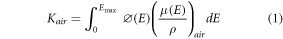
Where  (E) and Emax are the number of photons with energy E and the maximum value in the energy spectrum, respectively.
  is the mass energy absorption coefficient of photon with energy E derived from the NIST database (Hubbell and Seltzer 1996). Kair
was calculated by the
is the mass energy absorption coefficient of photon with energy E derived from the NIST database (Hubbell and Seltzer 1996). Kair
was calculated by the  obtained from the simulations when the detector volume was filled with air. The ratio of air kerma when there is a shield to that without any shield is taken as the air kerma transmission and is expressed as a percentage. It should be mentioned that energy spectrum of photons obtained in the detector was counted in 1 keV width energy bins and the corresponding coefficients were determined through interpolation from the values provided by NIST.
In the next step, for evaluation of the dose to organs at risk of a radiologist standing close to the x-ray tube and patient, a MIRD (Medical Internal Radiation Dose) phantom, i.e., a human male phantom of 173 cm height with various internal organs and material compositions was simulated. Simulated phantom is indicated in figures 3(A) and (B). The thyroid, skin, and gonads, owing to their proximity to the surface and heightened sensitivity to radiation, were designated as at-risk organs in the upper body, mid-body, and lower body regions, respectively. For calculation of skin dose, a skin cell of 5×5 cm2 area with 1.5 mm thickness (adopted from the work of Moradi et al 2017 on skin dose calculation) was considered. The radiation source was assumed as a point isotropic source located at 50 cm distance from the human phantom since this situation has been found as the worst-case scenario resulting in the highest dose to radiologist (Nowak et al 2020). The absorbed dose to organs at risks was calculated first without any shield (figure 3(B)) and then with an apron (with the height of 125 cm) covering the phantom upper body and the neck (figure 3(C)).

Figure 3. Designed human phantom (A) Interior organs of the phantom (B) Complete phantom (C) Phantom with the apron covering the upper body.
Download figure:
Standard image High-resolution imageLead aprons used in radiology are commonly of thicknesses of 0.25, 0.5 or 1 mm, while the minimum lead-equivalent thickness is considered 0.25-mm (Cheon et al 2018). Maximum protection is obtained by 1 mm thickness, however due to the weight issue, aprons have usually radiation shielding performance equal to 0.25–0.5 mm of lead (Nicol et al 2018). In order to have a comparison, in our simulation, aprons made of Pb, Ba5Ta4O15 without impurity and Ba5Ta4O15 with selected impurity were considered with 0.5- and 1-mm thicknesses.
3. Results and discussion
The results of the validation phase, which involved comparing the calculated mass attenuation coefficient ( ) from the Geant4 simulation with values obtained from the NIST database, are depicted in figure 4. The observed agreement confirms the accuracy of the implemented simulation model and the suitability of the geometry and setup.

Figure 4. Comparison between the mass attenuation coefficients of lead obtained from NIST with that calculated using the Geant4 simulations in this work.
Download figure:
Standard image High-resolution imageFigure 5 demonstrates photon spectra transmitted through various thicknesses of shield made of pure lead obtained from the simulations. Not surprisingly, the intensity of transmitted photons substantially decreases with increasing the shield thickness from 0.25 to 1 mm. As observed, with increasing shield thickness, lower energy photons are attenuated more pronouncedly and the peak of the spectrum shifts towards higher energies. As an initial evaluation, air kerma transmission in percentage for 1 mm thickness shield made of pure lead and barium tantalate was compared against shields composed of barium tantalate with 5% concentration by weight of different impurities. The findings from this test, as outlined in table 2, indicate that the introduction of 5% impurities does not improve the shield's attenuation capacity. However, there is an exception in the case of Samarium addition, where it reduces the air kerma transmission when compared to pure barium tantalate.
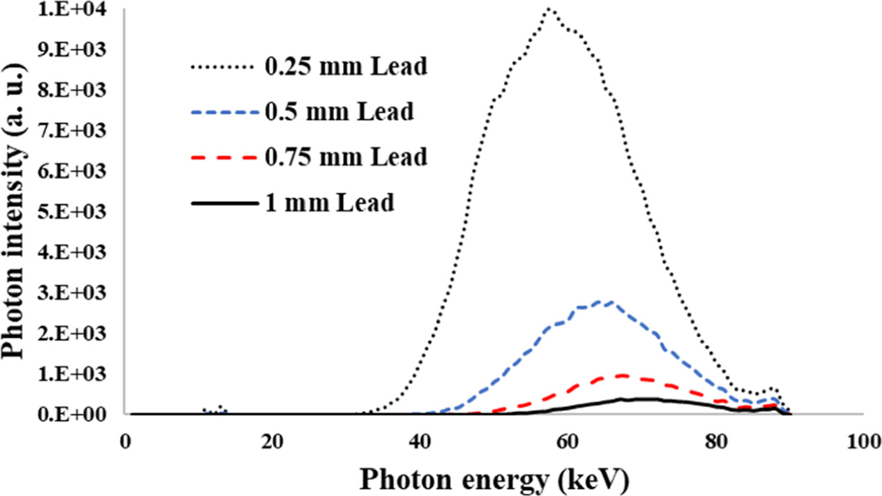
Figure 5. Spectra of photons transmitted through lead shield with thicknesses 0.25, 0.5, 0.75 and 1 mm.
Download figure:
Standard image High-resolution imageTable 2. Air kerma transmission calculated for 1 mm shield thickness. Data in this table corresponds to 5% by weight of various impurities in Ba5Ta4O15.
| Shield material | Air kerma transmission (%) | Shield material | Air kerma transmission (%) |
|---|---|---|---|
| Pb | 0.015 | Ba5Ta4O15 (Sm) | 0.021 |
| Ba5Ta4O15 | 0.023 | Ba5Ta4O15 (Tb) | 0.027 |
| Ba5Ta4O15 (La) | 0.031 | Ba5Ta4O15 (Dy) | 0.033 |
| Ba5Ta4O15 (Ce) | 0.030 | Ba5Ta4O15 (Ho) | 0.031 |
| Ba5Ta4O15 (Pr) | 0.033 | Ba5Ta4O15 (Er) | 0.032 |
| Ba5Ta4O15 (Nd) | 0.038 | Ba5Ta4O15 (Lu) | 0.027 |
| Ba5Ta4O15 (Gd) | 0.028 |
Following this, the simulations were conducted with various weight percentages of rare earth elements as 1, 2, 10, 12 and 15%. The results are presented as column charts in two figures 6 and 7 to be distinguishable. As depicted in figures 6 and 7, the air kerma transmission exhibits distinct trends with varying impurity concentrations, contingent on the specific impurity element. For certain elements like La, Ce, Sm, and Tb, the optimal weight percentage resulting in the lowest air kerma transmission appears to fall within the examined range of 1 to 15%. Conversely, for elements such as Dy and Er, an optimal concentration possibly exists beyond the studied range. Notably, the most effective shielding in terms of air kerma transmission was achieved with barium tantalate containing a 10% addition of Sm. It's essential to acknowledge that, in practical material production, lower concentrations, typically in the range of 1 to 10%, are more prevalent (Vani et al 2021, Jing et al 2023). Additionally, cost considerations for some of the investigated materials may limit the utilization of higher weight fractions in the composition. Consequently, concentrations exceeding 15% were not included in this study.
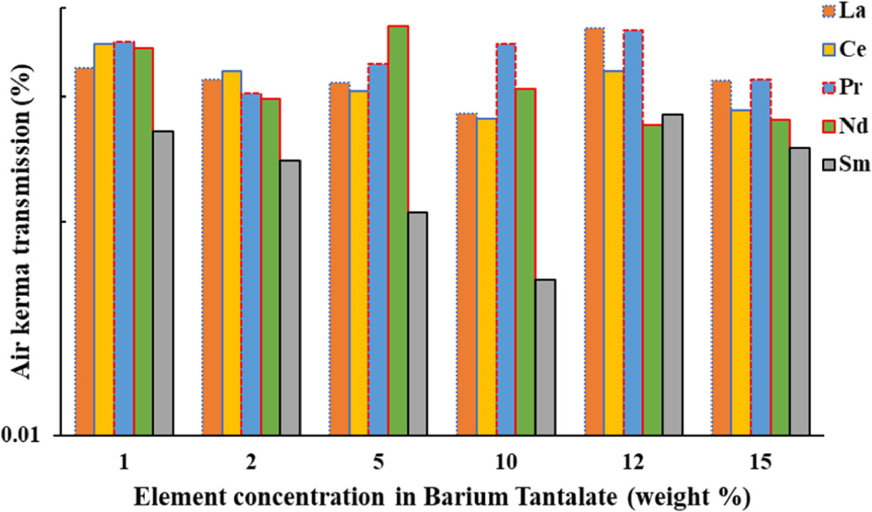
Figure 6. Air kerma transmission calculated for 1 mm thickness shield composed of Ba5Ta4O15 with various concentration of rare earth elements (La to Sm).
Download figure:
Standard image High-resolution image
Figure 7. Air kerma transmission calculated for 1 mm thickness shield composed of Ba5Ta4O15 with various concentration of rare earth elements (Gd to Lu).
Download figure:
Standard image High-resolution imageTo investigate variations in air kerma transmission as a function of shield thickness, we compared a lead shield with pure barium tantalate and barium tantalate containing 10% Sm impurity across a range of thicknesses from 0.25 to 1 mm. The findings of this assessment, illustrated in figure 8, clearly demonstrate that, when an equal-thickness shield is employed, Ba5Ta4O15 provides weaker protection compared to lead. However, the results also reveal an enhanced performance of Ba5Ta4O15 when it includes a 10% Sm impurity.

Figure 8. Air kerma transmission percentage through various thicknesses of different shield materials.
Download figure:
Standard image High-resolution imageGiven that the primary concern in interventional radiology shielding applications is the weight of the shield (apron), rather than just its thickness, we introduced a factor termed MDAKT. This factor, derived from the multiplication of mass density and air kerma transmission, facilitates a more meaningful comparison, considering both shield weight and radiation attenuation. MDAKT serves as an indicator for identifying compositions that not only exhibit radiation attenuation performance equivalent to lead shielding but also possess comparable or lower mass. The results for this parameter are detailed in table 3. Noting that MDAKT for 1 mm lead is  all compositions with equal or better shielding performance are highlighted in the table.
Table 3. Comparison of the parameter MDAKT (mass density × Air kerma transmission) calculated for 1 mm shield composed of Ba5Ta4O15 with different amount of various impurities.

|
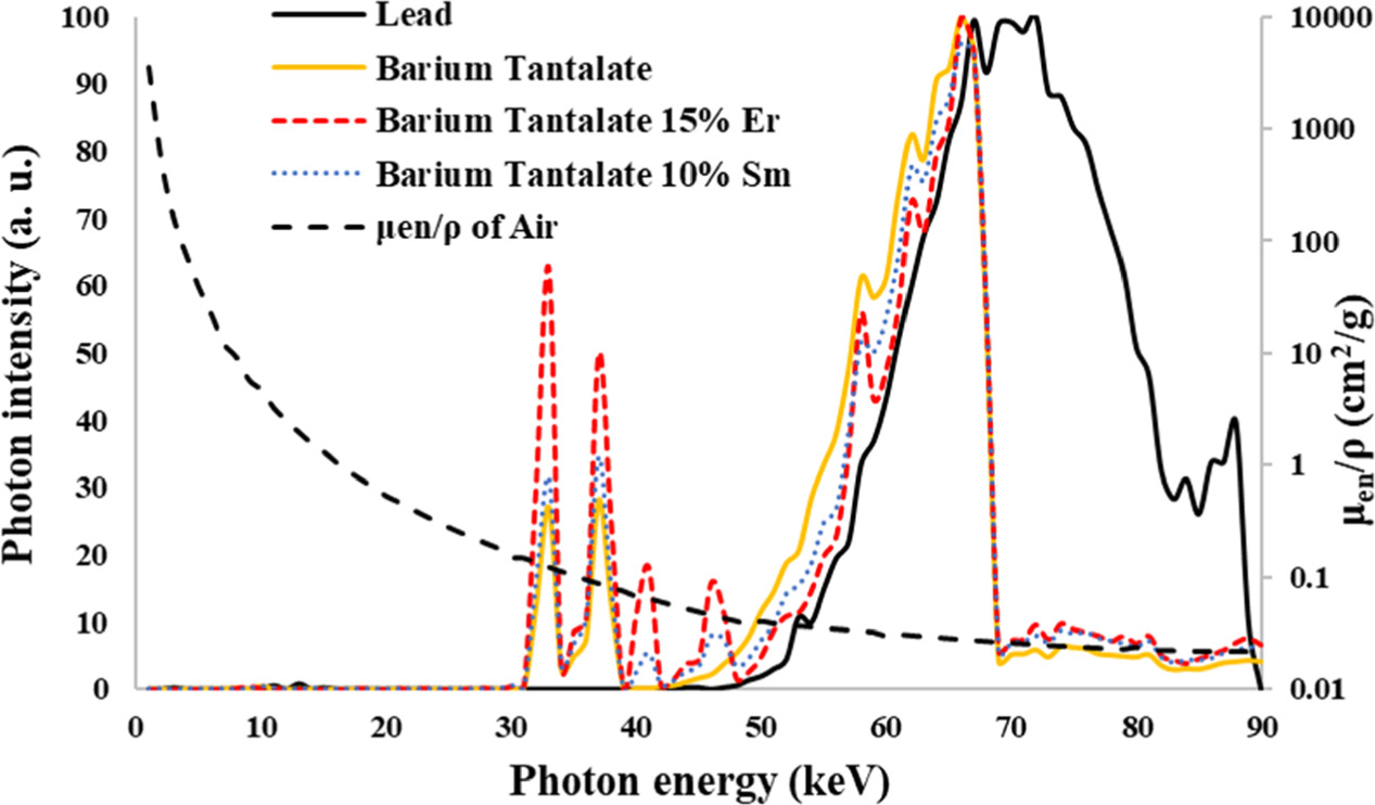
Compositions identified with better shielding performance are Ba5Ta4O15 containing dissimilar concentrations of Sm, Gd, Tb, Dy and Er ranging from 5 to 15% while the lowest transmission values obtained for Ba5Ta4O15 with 10% Sm and Ba5Ta4O15 with 15% of Er. It's important to bear in mind that, with cost-effectiveness in consideration, only the elements 62Sm, 64Gd and 68Er are economically feasible for routine shielding applications. Moving forward, we conducted simulations to determine the minimum required thickness of these two selected compositions to achieve an equivalent radiation protection level as that of 1 mm of lead (equivalent air kerma transmission). The results indicate that Ba5Ta4O15 with a 10% Sm impurity or 15% Er impurity requires only a slightly higher thickness, approximately 2% more (i.e., 1.02 mm), to attain the same air kerma transmission as 1 mm of lead. In case of Ba5Ta4O15(10% Sm) this is equal to over 31% reduction in weight ( ) while providing equal shielding performance. In these two instances, we illustrate the comparison of transmitted x-ray spectra with those of the lead shield and pure Ba5Ta4O15 in figure 9. The intensities in the spectra have all been normalized to their maximum values. It's important to note that the calculation of air kerma transmission involves multiplying the photon fluence within each energy bin by the corresponding mass energy absorption coefficient of air, represented as (
 ) of air. This value is also plotted on the secondary vertical axis for reference.
Figure 9. X-ray spectra of transmitted photons through 1 mm thickness lead shield and two barium tantalate compositions with equivalent air kerma transmission (The measure for the mass energy absorption coefficient of air is demonstrated on the secondary vertical axis on the right side).
Download figure:
Standard image High-resolution image
When comparing the spectra transmitted through lead and Ba5Ta4O15 compositions, a crucial observation is the absence of the portion of the spectrum with energies exceeding 67.42 keV in the case of Ba5Ta4O15 compositions. This phenomenon arises from the K-absorption edge of tantalum, situated precisely at 67.42 keV (while lead's K-edge is at 88 keV). Photoelectric absorption is the mechanism responsible for absorbing photons with energies surpassing the K-edge. Therefore, the presence of diverse elements in the shield composition leads to attenuation in various regions of the transmitted spectra. The influence of the Er and Sm K-edges, present in the composition, is also observable as declines in the continuous spectra of transmitted photons with energies exceeding 57.48 keV and 46.83 keV, corresponding to the Er and Sm K-edges. The results indicate that barium tantalate with a 10% Sm impurity provides the most effective attenuation for the studied x-ray spectrum. It should be noted, however, that while Ba5Ta4O15(10% Sm) exhibits lower attenuation (higher transmission) in the 50–67 keV energy range, it demonstrates higher attenuation than Ba5Ta4O15(15% Er) in the 30–50 keV range. Considering the fact that the mass energy absorption coefficient is higher in the lower energy range of photons and factoring in their respective densities (7.67 for Ba5Ta4O15(10% Sm) versus 7.90 for Ba5Ta4O15(15% Er)), the overall shielding performance of Ba5Ta4O15 (10% Sm) was determined to be superior.
In the final step, absorbed dose within the selected organs at risk was calculated first without the apron and then in a series of simulations with presence of 1 mm and 0.5 mm thickness aprons composed of lead and Ba5Ta4O15 containing 10% by weight of Sm. Er impurity was not analyzed in these simulations, however from the air kerma transmission outcomes, it is expected to produce similar results with a slightly higher shield mass. The calculation involved subtracting the dose delivered to organs at risk in the shielded scenario from that in the unshielded scenario, and the results are presented as a percentage reduction in tables 4 and 5. Additionally, the apron's mass was computed and is detailed in the tables. These findings indicate that within the examined photon energy range, barium tantalate containing 10% concentrations of Sm (as well as other elements as specified in table 3) can offer photon shielding properties on par with lead.
Table 4. Comparison of the shielding performance of 1 mm lead with 1 mm barium tantalate containing 10% by weight of Sm.
| Shield | Pb | Ba5Ta4O15 (10% Sm) |
|---|---|---|
| Mass (kg) | 7.45 | 5.14 |
| Reduction of dose to thyroid (%) | 98.82 | 98.72 |
| Reduction of dose to skin (%) | 99.11 | 99.03 |
| Reduction of dose to gonads (%) | 98.90 | 98.73 |
Table 5. Comparison of the shielding performance of 0.5 mm lead with 0.5 mm barium tantalate containing 10% by weight of Sm.
| Shield | Pb | Ba5Ta4O15 (10% Sm) |
|---|---|---|
| Mass (kg) | 3.82 | 2.64 |
| Reduction of dose to thyroid (%) | 97.76 | 97.71 |
| Reduction of dose to skin (%) | 98.18 | 98.24 |
| Reduction of dose to gonads (%) | 97.76 | 97.70 |
Regarding toxicity profile of compositions investigated in this study compared to lead shield, it should be noted that barium tantalate, particularly in the form of a composite containing cerium, is reported to be non-toxic (Jia et al 2016). Despite its widespread applications in electronics, crucial safety information is notably absent for barium tantalate in major chemical databases such as the Hazardous Substance Data Bank (HSDB), European Chemicals Agency (ECHA), or PubChem. This absence contrasts sharply with lead, for which hazardous statements are readily available. For instance, lead, as indicated in its safety data sheet, poses serious risks, including damage to the kidneys, blood-forming systems, central nervous system, and digestive tract through prolonged or repeated exposure. Moreover, lead is associated with potential harm to unborn children, breast-fed infants, suspected effects on fertility, suspected carcinogenicity, and harm to aquatic life. When considering lanthanides added to barium tantalate composition in this study individually, they are not highly toxic and are often used in various applications including medical imaging and technology, where their potential toxicity is carefully considered and managed. They are typically considered to have low toxicity levels. The available data suggests that barium tantalate, especially in composite forms containing selected rare earth elements offers a promising alternative with notably lower toxicity profile making it an attractive option for radiation protection applications where safety considerations are paramount. Discussed shield compositions offer notable advantages not only due to their non-toxic nature in contrast to lead but also because they are approximately 31% lighter in weight, as demonstrated above.
4. Conclusion
In this study, our primary objective was to assess the photon attenuation properties of barium tantalate incorporating varying concentrations of rare earth elements for its application in interventional radiology shielding. The accuracy of our simulation approach was initially validated through a comparison of mass attenuation coefficients with those obtained from a reference database, using lead as a benchmark. For an experimentally acquired x-ray spectrum, we calculated the air kerma transmission as a key indicator of the material's shielding effectiveness. Our results demonstrated that pure barium tantalate, without impurities, couldn't match the radiation shielding capabilities of lead. However, the addition of specified concentrations of Sm, Gd, Tb, Dy, and Er led to comparable or superior shielding performance while allowing for a significant reduction in shield mass. The most effective shielding in terms of air-kerma transmission was achieved using barium tantalate with 15% Er or 10% Sm impurities. Notably, the latter offered a reduction in absorbed dose to organs at risk in our human phantom model similar to that of a lead apron, while simultaneously reducing the apron's mass by 31%. The outcomes of this study hold significant importance in guiding future endeavors related to apron and shield production for use in radiation environments, particularly within interventional radiology settings. Moreover, the methodology employed in this research can be adapted for tailored shield optimization in specific radiological scenarios.
Data availability statement
All data that support the findings of this study are included within the article (and any supplementary files).
Conflict of interest
The authors declare that they have no conflicts of interest regarding publication of this manuscript.