
Abstract
In this work, we present an experiment conducted at the S-EBIT-I ion trap of GSI. It involved the study of ion-electron collisions of Fe and Ba ions in various charge states with the electron beam. Characteristic x-ray radiation emitted during the continuous interaction was recorded utilizing an energy-dispersive maXs-30 detector based on metallic-magnetic calorimeter (MMC) technology. Optimizations to the applied sensitivity-drift correction and energy calibration procedures significantly improved the achieved energy resolution compared to previous applications of a similar detector. This made it possible to individually resolve and identify overlapping x-ray lines of iron and barium in a wide spectral range. As a demonstration of the outstanding detector performance, we used the recorded spectral data to extract an estimate of the charge state distribution of Fe ions in the trap. This experiment campaign marks an important milestone in the ongoing effort to enable the deployment of MMC detectors for future high-precision measurements in fundamental physics experiments.
Export citation and abstract BibTeX RIS

Original content from this work may be used under the terms of the Creative Commons Attribution 4.0 license. Any further distribution of this work must maintain attribution to the author(s) and the title of the work, journal citation and DOI.
1. Introduction
The observation of cosmic x-ray radiation stemming from plasmas [1] has been a crucial tool for building our understanding of the origin of our Universe for a long time (see for example [2, 3]). However, the correct interpretation of photon spectra recorded by satellites and telescopes requires a detailed understanding and accurate models of the physics behind the participating interaction processes. Because of the sheer amount of possible ionic reaction partners, available data sets (like for example SPEX [4]) are often sparse and the theoretical description of multi-electron systems is difficult. Therefore, laboratory astrophysics is an indispensable testbed for atomic and nuclear physical interactions, as it allows us to probe atomic systems of interest in a controlled and accessible experiment environment (see, e.g. [5, 6]).
In common practice electron-beam ion-traps (EBITs) are often used as reliable and convenient ion sources. They provide a table-top solution for the preparation of well defined ion targets starting from neutral atoms of almost every element. Of particular interest, thereby, is iron, as it marks the energetically most favorable nucleus for both fusion and fission reactions. The observation of x-ray radiation stemming from the interaction of ions with the electron beam requires the use of energy- or wavelength-dispersive photon detectors. In the past, multiple high-precision measurements of different charge states of Fe ions have been performed using crystal spectrometers (see for example [7, 8]). They offer a very high energy resolution (typically below  ), however, usually also have a narrow bandwidth and low quantum efficiency (in the order of
  ). Thus, for experiments where, e.g. the comparison of intensities between multiple spectral features is important, semiconductor based spectrometers have been used instead (see for example [9]). Their quantum efficiency and spectral bandwidth are larger by several orders of magnitude compared to crystal spectrometers at the cost of loosing energy resolution, which for most semiconductor detectors amounts to a few
  .
.
X-ray spectrometers based on microcalorimeters are developing into a promising tool in fundamental physics research as they combine advantages of both conventional photon detection technologies. At very low operation temperatures, this is achieved by converting the absorbed energy of incident photons into a proportional rise in temperature within the typically small detector volume. Using different technologies, this change in thermal energy can then be measured with high precision and in a broad spectral range [10]. Early experiments deploying microcalorimeters to measure x-ray radiation stemming from an EBIT have been performed utilizing neutron transmutation doped Germanium detectors [11, 12]. In the past, other groups successfully used detectors, e.g. based on Si-thermistors [13] or more recently transition-edge sensors at EBITs [14].
Metallic-magnetic calorimeters (MMCs), like the maXs-series detectors developed in cooperation with the KIP [15–17], use the temperature dependent magnetization change of the paramagnetic sensor material in conjunction with highly sensitive SQUID magnetometers to determine the rise of temperature [18]. This method not only enables detectors with high linearity [19] but also with one of the highest intrinsic energy resolving powers  at
at  [20] achieved to date. Combined with a fast rise time of up to
  [21] - which, for example, is necessary in coincidence measurement schemes for background suppression - MMCs therefore are prime candidates for high precision measurements in fundamental atomic physics research. However, because of their high sensitivity, MMC detectors - like most low temperature detectors - are also susceptible to external noise like electromagnetic interference or mechanical vibrations leading to measurement artifacts like temperature dependent sensitivity drifts. In order to compensate for these effects, correction algorithms in software are necessary together with corresponding measurement procedures during the experiments. In order to develop and benchmark these methods, in the past, multiple experiment campaigns have been conducted on the campus of GSI/FAIR [22, 23].
In this work we present the results of a long term test using a maXs-detector at the S-EBIT-I ion trap of GSI [24]. The experiment setup as well as its components is presented in section 2. The description of the data analysis and our results in section 3 is followed by a short conclusion and outlook in section 4.
2. Experiment
The experiment presented in this work involved the observation of x-ray radiation emitted during the continuous interaction of Fe ions with a focused electron beam. Therefore, a MMC detector of the maXs-30 series was placed directly in front of one of the viewports of the S-EBIT-I ion trap (see figure 1). In the following, a short description of the components and setup of the experiment is given.

Figure 1. The two photos show the positioning of the MMC detector at the 90∘ viewport of the S-EBIT-I from different angles. The detector chip itself is mounted at the tip of the cool finger attached to its cryostat. Indicated are the electron beam direction in blue and the observation axis of the calorimeter into the trap in orange. The plastic cap covering the Be-window of the viewport was removed before the measurement.
Download figure:
Standard image High-resolution image2.1. Ion trap: S-EBIT-I
The S-EBIT-I (also known as Stockholm R-EBIT) was originally developed by the Physics and Technology LLC Institute in Livermore, USA [25]. In 2005/06 the trap was installed in a dedicated laboratory at the AlbaNova University Center in Stockholm, Sweden where it was further developed [26]. Since its transfer to the campus of the GSI Helmholtz-Center for Heavy-Ion Research in Darmstadt, Germany, the S-EBIT-I is in operation at its experiment place in the X4 cave. The vacuum chamber surrounding the trap can be pumped to reach a pressure of below  and the entire trapping area is shielded from ambient temperatures by an actively cooled heat shield at 60 K. The Helmholtz-coil pair used to focus the electron beam is operated at a temperature of 4 K using a pulse-tube operated cold-head. This way, a magnetic field of up to 3 T can be generated in the vicinity of the trap, which leads to an electron-beam diameter of around
  . At typical beam currents of 150 mA this corresponds to a beam current density in the order of 4 kA cm−2. The acceleration voltage, thereby, can be adjusted up to 30 kV. X-ray emissions resulting from the continuous interaction of the trapped ions with the electron beam can be observed through a total of eight viewports around the approximately 2 cm long ion-target. Overall, electron densities up to
  and ion densities of
  can be achieved [27]. The element to be measured can be brought into the system via a dedicated gas-injection system [26]. It allows the transfer of an adjustable amount of gas-atoms or -molecules from a pre-vacuum chamber through a needle valve directly into the electron beam. Simultaneously, a second gas of a lighter element can be mixed into the gas-flow. This enables the exploitation of evaporative cooling effects of the trapped ions by the lighter ions [28].
2.2. Detector: maXs-30
X-rays emitted during the ion-electron collisions were recorded using a maXs-30-type detector array with sensors based on MMC technology. A detailed description of the detector can be found in [17]. It has an active detection area of  divided into
  pixels (see figure 2). For stabilization of the signal baseline against temperature-drifts, two pixels each are connected to the same read-out and amplification stage in a gradiometric layout. The sensors' temperature-dependent change of magnetization in an external, static magnetic field is read out via inductively coupled dc-SQUIDs. A first amplification at low temperatures is applied to each of the signals utilizing a subsequent N-SQUID series array stage (see [18] for details). Finally, room temperature electronics further amplify the signal and provide a feedback signal to the read-out stage to fix the SQUID's operation point in a flux-locked loop (FLL). The design of the components is optimized for absorbers made from gold with around
  thickness, to have a quantum efficiency of 80% at a photon energy of
  and
and  below
below  [29]. The absorbers of the detector used in this experiment, had a thickness closer to
  , thus, slightly reducing its expected efficiency towards higher photon energies. However, in the spectral region of interest below
  , the impact on the quantum efficiency is negligible. Additionally, a smaller detector volume is expected to increase the signal-to-noise ratio of the calorimeter due to the fact that its sensitivity is inversely proportional to the thermal capacity of the detector and thus its overall volume. Under ideal conditions, an intrinsic energy resolution of about
  full width half maximum (FWHM) at
  and
and  in the low energy limit was reported for a comparable maXs-30-type detector (at 25 mK substrate temperature) [20]. The signal decay time of the utilized calorimeter was around 5 ms (time to reach the baseline level after the absorption of a photon). This is compatible with an average photon rate of around
  observed during the experiments. The operation of the detector requires substrate temperatures of below 30 mK to exploit its full potential. By utilizing a
  dilution cryostat a continuous measurement at operation temperatures down to
  is achievable. Furthermore, the detection of magnetic flux changes in fractions of the magnetic flux quantum requires a good shielding against external magnetic fields. This is particularly important for a setup in the vicinity of the Helmholtz-coil pair of the EBIT. Therefore, the maXs-30 detector uses a soft metallic alloy shield as well as a superconducting shield made from Niobium to cover the detector and the SQUID read-out and amplification stages.
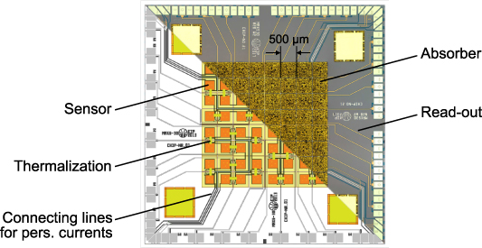
Figure 2. The image shows a microscopy image (upper right corner) of the maXs-30 detector utilized in the presented work, overlapped by its concept sketch (lower left corner, both images are taken from [17]). The connections labeled as 'Connecting lines for pers. currents' are used to prepare the persistent current for generating the required static magnetic field and 'Read-out'-lines connect the paramagnetic sensors to the SQUID read-out stage. Reproduced with permission from [17].
Download figure:
Standard image High-resolution image2.3. Setup and calibration
The microcalorimeter was set up in front of the 90∘ viewport of the EBIT with an air gap of  . The resulting distance of
  between the detector and the interaction area within the trap yields a total solid area coverage of 0.25 msr. Through the Be window of the EBIT and the x-ray entry window assembly of the maXs-30 (see [17] for details) a transmission of 65% of x-ray photons at
  is expected. During the measurement, Fe ions were supplied to the trap in the form of gaseous Ferrocene
 . By adjusting the anode voltage, several different electron beam currents between 11 mA and 25 mA were used throughout the measurement campaign. The acceleration voltage was fixed to 10 kV. In order to focus the electron beam, the Helmholtz-coil pair was supplied with a current of 17 A, resulting in a magnetic field strength of
  in the trapping region. To prevent the accumulation of heavier ions in the trap over time the measurement was performed in cycles. First, the trap was closed for 500 ms to allow the successive build-up of Fe-ions with increasing charge states due to Coulomb-ionization through the electron beam. Afterwards, the trap was briefly opened to extract all contained ions and to prepare it for the next trapping cycle.
To extract the measured photon energies from the MMC signals, a finite response filter-based approach was applied. The procedure closely follows the analysis scheme described in [30] and consists of a combination of a moving window deconvolution-filter followed by a moving average filter. For the energy calibration of the recorded x-ray data, a  and a
and a  source were positioned in front of the EBIT viewport opposite to the MMC detector, each at a time. At first, x-rays and γ-photons from the radioactive sources were observed through the EBIT alternating with the measurement periods. Later, the calibration was performed simultaneously with photons from the trap. The calibration itself was performed by fitting Gaussian distributions to the peaks of several calibration lines, in order to determine their position in the spectra. A second order polynomial was then used to fit the measured line positions to their well-known calibration energies (for 55Mn-Kα1, 55Mn-Kα2, 55Mn-Kβ1, 237Np-Lα1, 237Np-Lβ1, 237Np–
 , and 237Np–
 ) taken from literature [31].
The sensitivity of the MMC sensors is dependent on their substrate's temperature. Therefore, small fluctuation of the detector chip temperature (up to a few mK due to external vibrations of the cryostat) over time lead to corresponding variations of the measured energies, broadening the achieved instrumental resolution up to several  FWHM. This effect can be compensated by exploiting the fact, that asymmetries in the gradiometric pixel-pairs lead to a sensitivity of the signal baseline level to the substrate temperature as well [16, 17]. Although for most pixels this asymmetry is negligible, a dedicated pixel-pair with a deliberately imbalanced setup can be used as an on-chip thermometer. During the experiment, a temperature-drift correction procedure was implemented, that consisted of recording the baseline offset voltage of the temperature-sensitive pixel-pair for each triggered photon event. By performing a linear regression of the measured energies for events associated with the
  calibration line to the temperature-dependent offset values, a compensation curve was determined. Applying the compensation to all events then eliminates most sensitivity-drift effects and improves the detector resolution greatly. Due to occasionally occurring sudden shifts of the read-out SQUID electronics' operation point (most likely due to external magnetic noise), the recorded temperature reference changes accordingly. These discontinuities need to be identified in software as well and respective temperature corrections must be determined for each stable interval in between. After individually drift-correcting and calibrating the photon energies measured for each pixel, events from all pixels can be accumulated and used to calculate the combined spectrum.
3. Analysis of the data and results
The measurement campaign took place over the course of more than nine months. During the first runtime, the calibration and temperature-drift correction procedures were finalized and put to the test. First results of the experiment can be found in [24]. Because of a facility-wide power outage the EBIT was severely damaged and had to be repaired. Afterwards, a second runtime followed during which the EBIT was set back up.
3.1. First run
In the first runtime, an x-ray spectrum only containing transitions from Fe-ions was recorded, which can be seen in figure 3. During the interaction of the ions with the electron beam, electron impact excitation and ionization leads to the formation of K-shell vacancies leaving the ions in an excited state. The shown spectrum contains Kα photons emitted during the subsequent relaxation of an L-shell electron into the ground state. For neutral iron the Kα transition energy amounts to  [32] which coincides with the low-energetic boundary of the measured spectral distribution. By removing K- and L-shell electrons from the ions, their shielding effect on the nuclear Coulomb potential acting on the remaining electrons decreases. Therefore, the binding energy and, consequently, the transition energy between the bound states increases. The literature value for the ionization energy of Fe24+ is given as
  and
and  for Fe25+ [33]. Thus, in principle, Fe ions could be fully ionized in collisions with an electron beam with
  beam energy. At the same time, electrons from the beam are also being captured by the positively charged ions. The charge state distribution of the trapped ions therefore results in a time dependent equilibrium between ionization and radiative recombination (RR) [34] processes. It is defined by the electron beam-energy, and -density, as well as the target density and interaction time (see [35] for a detailed description of the dynamics). An approximation of the reaction cross sections using the flexible atomic code (FAC) [36] suggests that the removal of a L-shell electron is slightly more probable than the recombination for the given experiment parameters. The resulting accumulation of ions with increasing charge states over time matches the observed spectral behavior. Ionizing a K-shell electron, however, is two orders of magnitude less probable than the L-RR. After removing all L-shell electrons, it is significantly more probable for the ions to recombine with another electron than to remove a K-shell electron. Therefore, no charge states beyond Fe24+ are expected to occur. A comparison of the Kα energy for He-like iron of
  [8] with the high-energy boundary of the recorded spectrum agrees with this conclusion. During the experiment, a beam current of 11 mA was set. Based on the association of their most prominent transitions to the corresponding spectral features, the centroid position of the charge state distribution is approximated to fall between 18+ to 20+. The influence of the M-shell electrons on the binding energy of K- and L-shell electrons is weak. Thus, the change of Kα transition energies for charge states beyond neon-like iron is expected to vary significantly less. During the first run, the calibration utilizing the radioactive sources was performed in a brief periods before the EBIT measurements themselves. Due to changing read-out conditions (as explained in section 2.3), the sensitivity-drift behavior of the MMC sensors changed between measurement and calibration steps. Therefore, a direct correlation between the recorded temperatures and measured energies was difficult and remaining uncompensated sensitivity-drift lead to an overall instrumental resolution of
  FWHM at
  . Compared to the expected
  FWHM from previous measurements under ideal conditions, this proves the necessity of proper temperature correction procedures. The peak of superposed Kα transitions below
  stemming from neutral iron up to Fe16+ could not be resolved.

Figure 3. The spectrum contains x-rays stemming from Kα transitions in different charge states of iron. The charge state labels are associated to their most prominent transition and aim to give a rough orientation of the most significant spectral features. The Kα transition energies for neutral [32] and He-like iron [8] are highlighted.
Download figure:
Standard image High-resolution image3.2. Second run
After the EBIT was damaged during a facility-wide power outage, it had to be disassembled, cleaned, repaired, and rebuild. During the setup phase of the reassembled EBIT, more x-ray photons were recorded in a spectrum shown in figure 4. Besides the expected iron Kα transitions it also contains several lines stemming from Ba ions. Barium is present inside of the trapping volume, because of the barium-oxide coating covering the cathode (used to improve electron emission characteristics). Recording the baseline voltage of the temperature-sensitive pixel-pair for every photon event and the simultaneous calibration using the
  source during the entire measurement period made an efficient temperature-drift correction in software possible (as described in section 2.3), leading to a significant improvement of the achieved energy resolution to
  FWHM at
  (single pixel resolutions down to
  ). This corresponds to a doubling of the achieved resolving power compared to the first run. Furthermore, temperature-drift related spectral artifacts as observed in previous measurements utilizing a maXs-30-type detector [23], could be suppressed entirely. The remaining uncorrected temperature drift effects still make up most of the difference to the measurement under ideal conditions (
 FWHM). The advantages of using a MMC detector becomes particularly obvious when comparing its spectrum with one, that was recorded by a conventional Si(Pin)-diode (XR-100CR by Amptec Inc.). The diode was also attached to one of the EBIT viewports and recorded x-rays at the same time as the MMC. However, in contrast to the results yielded by the semiconductor detector, spectral features from different charge states of Ba and Fe can be individually resolved utilizing the calorimeter based spectrometer. Using estimations for their transition energy from model calculations performed in FAC, transitions from the M- to the L-shell of Barium (see figure 5 top), as well as from the N- and O- to the L-shell (see figure 5 bottom) were identified. Due to the increase of ionization energy from
  for Ba45+ to
  for Ba46+ [33], FAC calculations of the corresponding cross sections suggest a similar imbalance between the M-RR and L-shell ionization as discussed for iron in section 3.1. This matches the observation that neon-like barium (Ba46+) is the highest charge state present in the spectrum, despite that fact that the production of Ba51+ was energetically possible using
  electrons.

Figure 4. The spectrum gives an overview over all x-ray events recorded during the second S-EBIT-I measurement period. It contains lines from both Ba as well as Fe ions (grouped and labeled by the shells of their involved initial and final states, e.g., transitions from the M- to the L-shell). The comparison between the Si(Pin)-diode based semiconductor detector (red) and the MMC spectrum (blue) highlights the significant difference in achievable energy resolution between the technologies. Note, that the Si(Pin) spectrum was scaled down to match the peak intensities measured by the maXs-30 for better comparison. Additionally, characteristic x-ray radiation from copper and zinc is visible between 8 and  . These peaks can be explained by x-ray fluorescence of a brass aperture in front of the detector chip, resulting from an excitation by γ-photons stemming from the 241Am-calibration source.
Download figure:
Standard image High-resolution image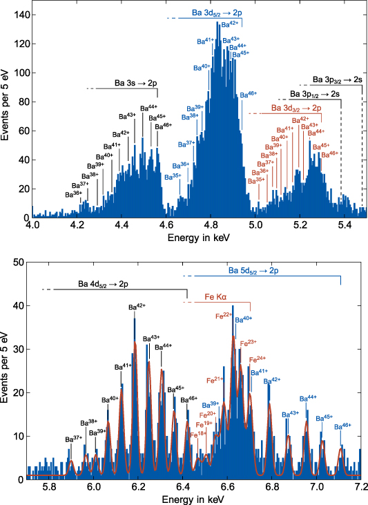
Figure 5. The two spectra show details of transitions form the M- to the L-shell in different charge states of Barium ions (top), as well as from the N- and O- to the L-shell (bottom) recorded at the EBIT. The association of charge states to peaks was accomplished by comparing their corresponding transition energies with values estimated by FAC. The bottom spectrum also contains x-ray events stemming from Fe Kα transitions. A fit to a model containing transition energies from a previous measurement of Fe [8] and all calculations for Ba was performed (red line).
Download figure:
Standard image High-resolution image3.3. Charge state distribution
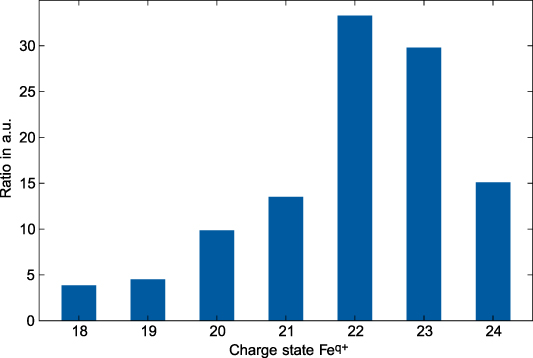
In order to estimate the charge state distribution of iron ions in the trap, a fit to a model function was performed. The model consists of a superposition of Gaussian peaks, with their mean energy fixed to the identified transition energies calculated for barium and previously measured for iron ions [8]. A least squares minimization to the spectral data yields the amplitudes of the individual x-ray lines. All peak heights for transitions belonging to the individual charge states are then accumulated and used as a rough qualitative measure for the abundance of that state. The resulting distribution can be seen in figure 6. It was later discoverd, that after the rebuild of the EBIT, electrical discharges between internal components lead to a feedback of voltage spikes into the surrounding electronics. This caused the controller responsible for the trap timing cycle to temporarily malfunction. As a result, the trap was closed significantly longer than expected. On the one hand, this explains why barium ions, which are around 2.5 times heavier than iron ions, were accumulated inside of the trap in the first place. On the other hand, it matches the observation that the centroid of the Fe ion's charge state distribution is shifted towards higher values compared to the first run.
Figure 6. Displayed is the rough, qualitatively estimated charge state distribution of iron ions in the EBIT during the second run time. It was derived by fitting a model with corresponding transition energies to the x-ray spectrum recorded by the maXs-30 calorimeter.
Download figure:
Standard image High-resolution image
4. Conclusion and outlook
During the S-EBIT-I measurement campaign, the maXs-30 detector and its cryostat were reliably operational for more than nine months, with only few and short maintenance down times in between. Active work at the EBIT meant, that the MMC was constantly subject to mechanical vibrations and changing temperatures of the surrounding area, while it was measuring x-ray photons. However, because of the optimized sensitivity-drift correction- and calibration-methods developed during the experiment, the achieved energy resolution improved by a factor of two compared to past applications [23]. Particularly, previously observed artifact peaks resulting from unstable operation points were successfully eliminated by implementing the continuous sensitivity-drift compensation during the entire measurement period.
Utilizing the outstanding performance of the MMC detector, superposed x-ray transitions stemming from a wide range of charge states of both barium and iron ions could be individually resolved and identified. As a demonstration, an estimation for the charge state distribution of iron ions in the trap was successfully extracted from the measured overlapping Ba Lγ and Fe Kα peak intensities. Overall, despite being placed in direct vicinity to the magnetic field of the superconducting EBIT magnet, the MMC-based spectrometer performed almost to its design specifications. This marks an important milestone for the future deployment of maXs-series detectors, e.g., at accelerator beam lines, where typically access to the experiment setup during the beam time is limited and external sources of strong noise like pumps and beam magnets are present. Together with the coincidence-based background suppression technique demonstrated with a maXs-100 detector during a recently performed beam time at CRYRING@ESR of GSI [37], maXs-series x-ray spectrometers have come a long way towards becoming an indispensable tool for high-precision measurements in fundamental physics research.
Moreover, the presented results underline the unique research potential provided by EBIT ion sources in combination with micro-calorimeter and in particular with MMC detectors. The high spectral bandwidth combined with the high energy resolution seems to be an ideal tool for investigating the often very complex x-ray emission in experiments with EBIT sources and allows the identification of lines that in the past were often hindered by line blends due to the comparatively low resolving power of conventional solid state detectors. Currently, a maXs-series detector is being set up in the newly available dedicated laboratory at the Helmholtz Institute Jena. In addition, the utilization of a PolarX-EBIT [38] as a local ion target for ion-electron-collision measurements is under discussion. Beside the relevance of such studies for future detailed investigations of electron beam driven collision studies with highly-charged ions, this combination of an EBIT with an MMC detector would provide an excellent test laboratory to further refine and improve the operation of MMC detectors under realistic experiment conditions. Planned enhancements, on the one hand, include the reduction of the detector's susceptibility to external sources of noise, e.g. by dampening vibrations of the cryostat. On the other hand, the acquisition of more information about the detector's environment is necessary. For example, the usage of multiple temperature sensitive pixels, which has been implemented recently, gives rise to a more effective software-based compensation of artifacts induced by sensitivity-drift effects. In particular for the preparation of future experiment campaigns at accelerators, such a test and development platform would therefore be highly beneficial.
Acknowledgments
We thank all participating members of the research division for atomic, quantum and fundamental research of GSI and of the KIP that helped with the experiment setup and execution. This work was created within the SPARC collaboration. We acknowledge financial support by the European Union and the federal state of Thuringia via Thüringer Aufbaubank within the ESF project (2015 FGR 0094).
Data availability statement
The data cannot be made publicly available upon publication because they are not available in a format that is sufficiently accessible or reusable by other researchers. The data that support the findings of this study are available upon reasonable request from the authors.