Case Report

The Potential of Integrative Cancer Treatment Using Melatonin and the Challenge of Heterogeneity in Population-Based Studies: A Case Report of Colon Cancer and a Literature Review

by
Eugeniy Smorodin
1,*,
Valentin Chuzmarov
2 and
Toomas Veidebaum
1
1
Department of Chronic Diseases, National Institute for Health Development, Paldiski mnt 80, 10617 Tallinn, Estonia
2
2nd Surgery Department, General Surgery and Oncology Surgery Centre, North Estonia Medical Centre, J. Sütiste Str. 19, 13419 Tallinn, Estonia
*
Author to whom correspondence should be addressed.
Curr. Oncol. 2024, 31(4), 1994-2023; https://doi.org/10.3390/curroncol31040149
Submission received: 28 January 2024 / Revised: 19 March 2024 / Accepted: 27 March 2024 / Published: 3 April 2024
(This article belongs to the Section Gastrointestinal Oncology)

Abstract

:
Melatonin is a multifunctional hormone regulator that maintains homeostasis through circadian rhythms, and desynchronization of these rhythms can lead to gastrointestinal disorders and increase the risk of cancer. Preliminary clinical studies have shown that exogenous melatonin alleviates the harmful effects of anticancer therapy and improves quality of life, but the results are still inconclusive due to the heterogeneity of the studies. A personalized approach to testing clinical parameters and response to integrative treatment with nontoxic and bioavailable melatonin in patient-centered N-of-1 studies deserves greater attention. This clinical case of colon cancer analyzes and discusses the tumor pathology, the adverse effects of chemotherapy, and the dynamics of markers of inflammation (NLR, LMR, and PLR ratios), tumors (CEA, CA 19-9, and PSA), and hemostasis (D-dimer and activated partial thromboplastin time). The patient took melatonin during and after chemotherapy, nutrients (zinc, selenium, vitamin D, green tea, and taxifolin), and aspirin after chemotherapy. The patient’s PSA levels decreased during CT combined with melatonin (19 mg/day), and melatonin normalized inflammatory markers and alleviated symptoms of polyneuropathy but did not help with thrombocytopenia. The results are analyzed and discussed in the context of the literature on oncostatic and systemic effects, alleviating therapy-mediated adverse effects, association with survival, and N-of-1 studies.

1. Introduction

The hallmarks of cancer are genetic deregulation and epigenetic modifications caused by external and internal factors, which lead to abnormal cell growth and dissemination. A growing tumor creates a surrounding vasculature necessary for nutrition under conditions of hypoxia and deregulated metabolism and interacts with other cells in the microenvironment [1]. High heterogeneity, plasticity, and self-renewal help tumor cells avoid immune surveillance, become resistant to chemotherapy (CT) and apoptosis, and spread, creating metastatic niches. A developing tumor has both local and systemic effects on the body, which leads to cachexia, psychoneuroendocrine and immunoinflammatory imbalances, pro-tumoral cytokine production, aberrant myelopoiesis, immunosuppression, chronic inflammation, paraneoplastic syndromes, hemostatic abnormalities, and other disorders [2,3,4,5,6].
Due to the unsatisfactory efficacy and adverse effects of conventional therapies in advanced cancer, some patients themselves use additional or alternative treatments that can be beneficial, not helpful, or harmful. A patient’s response to treatment depends on the characteristics of the tumor and the patient’s state of health. Poor sleep, an unbalanced diet, distress, and lack of physical activity can significantly affect outcomes and life expectancy. All these factors can be improved by a patient through self-management, following the recommendations of oncologists and healthcare experts. In this regard, an integrated approach to treatment and improvement in overall health and quality of life is becoming increasingly important for personalized oncology [7]. Despite advances in conventional treatment, current therapies are insufficient to control metastatic disease. Colorectal cancer (CRC) continues to be the third-most diagnosed type of cancer but second in terms of mortality in 2020 [8]. Acquired resistance of tumors to CT, the harmful effects of CT and radiation therapy (RT), and the high cost and limitations of targeted therapy and immunotherapy (IT) stimulate the search for new treatment strategies [9,10,11,12]. To improve patient outcomes, a therapeutic approach using a combination of natural products that can simultaneously act on various pathways of tumorigenesis is attracting attention [13].
Melatonin (Mel) is involved in many vital cellular functions and affects the epigenetic mechanisms, gene expression, and signaling pathways associated with the emergence and progression of cancer. Mel is an immunomodulator and one of the transcriptional modulators affecting tumor homeostasis [14,15]. Mel, nutrients (zinc, selenium, and vitamin D), and plant polyphenols may be useful supplements in the treatment of cancer and require additional clinical trials [16,17,18,19,20], but the methods and doses used in many preclinical studies are inadequate for translation to clinical settings. Parallel randomized and placebo-controlled trials (RCTs) have encountered obstacles to resolving the contradictory results of adjunctive treatments due to the heterogeneity associated with many factors, including poor patient stratification, individual response to treatment, bioavailability, and the complex multifaceted relationship between a tumor and homeostasis. A personalized approach based on testing clinical parameters in response to integrative treatment in dynamic, patient-centered N-of-1 studies deserves greater attention. Integrative treatment with Mel directed to systemic effects in cancer and adverse consequences of conventional therapy may improve quality of life and prolong overall survival, as shown in some preliminary clinical studies discussed below. The present paper describes the dynamics of the clinical parameters of a patient with colon cancer who took Mel, nutrients, and aspirin. Integrative treatments are analyzed and discussed in the context of a review of the literature examining their oncostatic and systemic effects as well as the influence on conventional therapy and survival. Monitoring of clinical parameters before, during, and after CT with Mel as well as their comparison when taking Mel and its withdrawal may be considered an example for N-of-1 study designs of personalized responses to integrative treatment with Mel.

2. Materials and Methods

This analysis and monitoring of clinical parameters were based on patient protocols and laboratory tests at the North Estonia Medical Centre. An integrative treatment with Mel and nutrients was applied by the patient on his own initiative, and the design was based on the scientific literature and his qualifications and competence (Ph.D., biochemistry, pharmacology, and oncology). Doses of Mel and the period of its administration and withdrawal were selected by the patient himself. Investigations were carried out following the rules of the Declaration of Helsinki and in accordance with the ICH GCP Standards. The literature review was compiled using information from PubMed, Web of Science, Google Scholar, and Crossref. Based on the results of the completed study, this paper was written taking into account the patient’s consent.

3. Results

3.1. Colonoscopy, Computed Tomography, and Histology

A 69-year-old nonsmoking man was diagnosed with a malignant tumor of the sigmoid colon and two polyps during colonoscopy. His parents had no history of gastrointestinal cancer but were diagnosed with ovarian and prostate cancer. In July 2018, he was operated on. The distal part of the colon with a tumor along with the surrounding lymph nodes was removed. Tubular adenocarcinoma with tubular adenoma and hyperplastic polyps were found in biopsies and resected tumors. In addition, subserous tumor deposits in adipose tissue were found in the specimens. The tumor was classified as pT3N1c (Nx) M0 G2, stage III B. Immunohistochemical analysis revealed 80% for Ki-67 and 90% for p53. Metastases in the lung, liver, and abdominal cavity were not detected in preoperative computed tomography and twice after surgery for 4 years. During a third colonoscopy, one hyperplastic polyp was removed.
In cancer pathology, tumor deposits are defined as focal aggregates of cancer nodules located in the pericolonic or perirectal adipose tissue or adjacent mesentery separately from the invasive margin and lymph node tissue. The presence of tumor deposits is an independent unfavorable prognostic factor for patients with sigmoid colon cancer. Adjuvant CT can significantly improve survival in patients with subcategory N1c colon cancer [21,22].
Ki-67 is a nuclear protein associated with cell proliferation. A high percentage of Ki-67-expressing cells in the tumor material is an unfavorable prognostic factor for CRC patients [23]. However, the prognostic role of Ki67 in CRC remains controversial, which can be explained by a better response to adjuvant therapy in patients with a high tumor cell proliferation index. Rapidly proliferating cancer cells may be more vulnerable to adjuvant therapy. A favorable prognosis has been reported in patients having tumors with higher Ki-67 expression who underwent both surgery and adjuvant radiochemotherapy but not in patients who underwent surgery alone [24,25].
The p53 regulatory protein is a transcription factor that acts as a tumor suppressor. It can induce cell cycle arrest to repair DNA damage, induce apoptosis, block proliferation, and inhibit tumor angiogenesis. In addition, p53 is activated in response to stress factors, and it regulates various pathways inherent in oncogenesis. The TP53 gene is the most frequently mutated gene (>50% in human cancer cells). In general, patients with abnormal p53 have an increased risk of death both when assessing p53 protein accumulation and when analyzing mutations [26]. Mutations in TP53 are more likely to be associated with the risk of metastasis than immunohistochemically assessed accumulation of p53 protein in the primary tumor. However, mutations in TP53 can have different effects, including gain or loss of function, and affect the sensitivity of tumors to treatment in different ways [26]. CT and RT can also induce different mutations in tumor cells. Abnormal accumulation of p53 protein in tumors may also have a complex relationship with the tumor response to cytotoxic drugs and radiation. This may be one of the reasons for the contradictory results in studies regarding the benefit of CT in patients with overexpression of the p53 protein [26,27,28,29].

3.2. Systemic Chemotherapy

Eight cycles of CT were prescribed, but due to thrombocytopenia, four cycles of CAPOX with oxaliplatin (100–130 mg/m2) and capecitabine (2–3 g per day) along with antiemetics were performed. Adverse effects were observed, such as laryngopharyngeal dysfunction, numbness and tingling in the hands that occur with cold exposure, diarrhea, nausea, anemia, thrombocytopenia, and neutropenia. The adverse effects of CT were temporary and gradually disappeared. CT was suspended after the first cycle due to sinusitis and bronchitis, which were treated with antibiotics. Oral amoxicillin (2 g/day, 7 days) was ineffective; therefore, azithromycin (250 mg/day, 5 days) was administered orally, which was more effective.
CT with oxaliplatin/capecitabine causes neutropenia, lymphocytopenia, and thrombocytopenia [30]. Oxaliplatin is one of the most neurotoxic anticancer drugs, causing long-term peripheral neuropathy [31]. After completion of CT, the patient did not have any residual manifestations of neuropathy. Two months after CT, numbness and decreased sensation were noted in the toes and soles. Melatonin can promote neuritogenesis in oxaliplatin-challenged cells but does not interfere with the cytotoxic activity of oxaliplatin in a human colon cancer cell line; it can also reduce neuropathic manifestations [32,33,34]. The use of Mel or its combination with zinc [35] during and after chemotherapy could reduced and delayed the onset of CT-mediated neurological adverse effects in the patient.

3.3. Clinical Manifestations in Using Melatonin

The patient used a sublingual form of Mel (Melatosell Strong, Hankintatukku OY, Karkkila, Finland) in the evening and at night during the first awakening for 11 months, including doses of 1.9–5.7 mg/day a month before surgery and 5.7–7.6 mg/day after surgery. He took a dose of 19 mg/day during CT and 9.5 mg/day between cycles of CT. After a 1-year break, the patient resumed taking Mel at lower doses (1.9–3.8 mg/day) at 1-month intervals, and then at 1–2-week intervals. According to his subjective assessment, mild to moderate effects, such as sedation, intermittent sleep, and daytime drowsiness were noted. The use of Mel after diagnosis promoted relaxation, alleviated stress, anxiety, and depressive symptoms, thereby promoting falling asleep and longer sleep. Mel withdrawal did not cause insomnia or addiction.

3.4. Aspirin, Food Supplements, and Nutrition

The patient started taking low-dose aspirin (Hjertemagnyl, Takeda, Oranienburg, Germany, 75 mg daily) 8 months after surgery, according to its benefits and harms in an updated modeling study and results of a systematic review and meta-analysis [36,37]. The patient, for a long time before and after diagnosis, took selenium (Se) and zinc supplements at a daily dose of 50 μg of Se (selenomethionine and sodium selenite) and 10–15 mg of zinc. In addition, he took manganese, magnesium, and vitamins D3, B6, and C in generally accepted doses. Magnesium and B6 were used to reduce muscle cramps at night. The daily dose of Se was temporarily increased to 200 μg/day in the last cycle of CT and for 6 weeks after CT. The serum level of Se also showed an increase from 121 to 150 µg/L (analysis in Synlab). Optimal plasma Se values have been estimated at 90–120 µg/L [38], but higher Se levels might be associated with a reduced risk of certain cancers and better survival in those with CRC [39,40]. The patient was taking vitamin D3 (600–2500 IU) to maintain normal plasma levels of its metabolite calcitriol, which ranged from 63 to 104 nM. In addition, he took probiotics and prebiotics to normalize bowel function as well as natural polyphenols (after chemotherapy, periodically, with an interval of 1 month), namely, taxifolin (dihydroquercetin, 25 mg) and 600 mg of green tea extract (Evalar). The tablets were stored in a minimum volume of olive oil at 4 °C to protect against oxidation and improve the bioavailability of polyphenols [41].
The patient adhered to an active lifestyle and rational nutrition, according to beneficial recommendations [42,43,44,45,46,47]. He excluded roast, red, and processed meats, reduced the consumption of fatty and sweet foods, granting preference to dairy products, yogurts, fish, nuts, cereals, vegetables, fruits, olive and flaxseed oils, fish oil, and dietary fibers. The patient did not use other seed oils with an increased content of proinflammatory and pro-tumorigenic linoleic acid (omega-6 polyunsaturated fatty acid) [48,49,50,51]. He consumed, in increased amounts, berries containing polyphenols (bilberry, cranberry, etc.), green tea, and ginger, while alcohol consumption was kept to a minimum. A low body mass index may be a significant negative predictor of outcome in patients with CRC [52]. The patient lost 10% of his weight and, despite a normal appetite, did not regain his original weight. Taking melatonin may influence body weight [53].
Integrative treatment using dietary supplements and antioxidants can reduce the toxic and harmful effects of CT and RT on the body. On the other hand, together with conventional therapy, they can affect the effectiveness of therapy by removing therapy-induced cytotoxic reactive oxygen species (ROS) and influencing drug metabolism [54,55,56]. Opinions on the benefits or risks of antioxidants vary, and it is difficult to assess the overall outcome [54,57]. The category of “antioxidants” studied in reviews and meta-analyses includes substances with different mechanisms of action on normal and tumor cells, which are not limited to ROS removal. For example, polyphenols are being investigated as epidrugs that affect the epigenome program, thereby restoring cellular memory in cancer [1]. The antioxidant and prooxidant effects of Mel on human cell lines have been shown, depending on the concentration and duration of exposure. Mel can promote the formation of ROS and the death of some cancer cells as well as enhance the cytotoxic effect of drugs on tumor cells [58].

3.5. Clinical Parameters and Their Dynamics

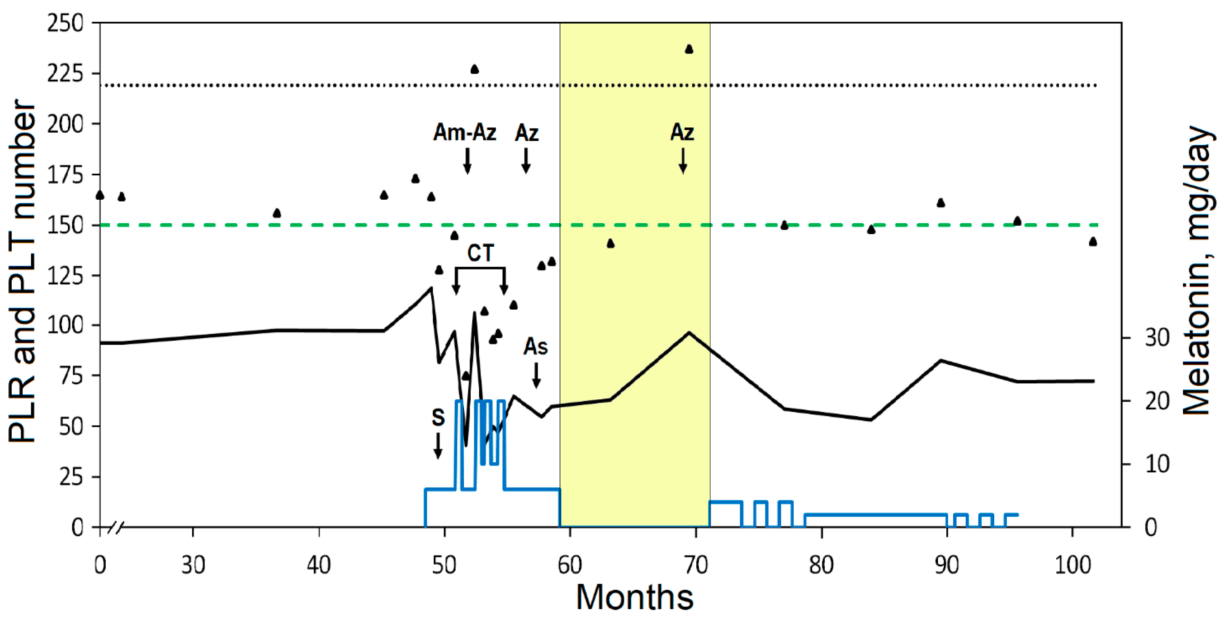
3.5.1. Tumor-Associated Systemic Inflammation

Systemic and local inflammation are considered as key elements in the development of cancer since inflammation drives tumor initiation, growth, progression, and spread [4,59,60]. Numerous studies collected in reviews [61,62] demonstrate that routinely used biomarkers associated with inflammation, namely, the ratio of neutrophils/lymphocytes (NLR), lymphocytes/monocytes (LMR), and platelets/lymphocytes (PLR), can be applied for prognostic CRC assessment. The risk of mortality has been found to increase in patients with a higher NLR and PLR and a lower LMR. This association can be considered as unfavorable systemic inflammation and, in terms of immunosuppression, causes a decrease in the lymphocyte population [4,60,63,64,65].
Reported values of prognostic sensitivity and specificity can be assessed as moderate or high, but the cut-off values and AUC (area under the curve) vary [61,62]. In addition, values of the ratios require unification since they are influenced by tumor localization, pathological data, and concomitant non-cancer diseases.
Postoperative inflammation-based prognostic markers can more accurately predict overall survival and recurrence-free survival in patients with stage III CRC [66]. The dynamics, association with survival, and clinical relevance of the ratios were studied by Herold et al. [67]. The authors note that personalized temporal changes in clinical parameters may more accurately reflect the risks of cancer progression. Furthermore, based on a prognosis-related blood cell count, an integrated prognostic inflammatory index with optimal cut-offs and nomograms was developed for personalized prediction of CRC survival [68].
The patient’s follow-up study showed that the preoperative NLR was slightly above the cut-off but decreased significantly after surgery and remained below the cut-off. The low preoperative LMR varied after surgery and during CT and then increased and remained above the cut-off (Figure 1). The low value of PLR associated with a low platelet (PLT) count remained below the cut-off (Figure 2). The cut-offs presented in Figure 1 and Figure 2 were selected based on the study with largest number of participants (5336) and with the highest proportion of patients with stage III CRC (48.3%) compared to other similar studies [69]. CT caused transient neutropenia, while the count of lymphocytes remained within the reference range. Notably, the reduced count of lymphocytes gradually increased after surgery and even during and after CT (Table 1). This was reflected in the lower NLR and higher LMR and may be related to the beneficial effect of Mel. Preliminary clinical studies have shown that the use of Mel can increase the LMR value that can potentially be used in antitumor IT [70,71]. A low lymphocyte count during chemotherapy is an unfavorable factor as it may predict worse disease-free survival in patients with colon cancer [72]. Tumor infiltration of T lymphocytes is an independent informative prognostic factor that was confirmed in a systematic review and meta-analysis of patients with CRC [73].
Mild anemia and preoperative eosinopenia were observed in the patient. Anemia was not associated with Mel use. The number of eosinophils significantly increased after surgery and remained within the reference range (Table 1). A decrease in the number of circulating eosinophils in patients with CRC has been associated with a shorter overall and disease-free survival [74]. Based on the prognostic value of eosinophil- and basophil-related markers, a scoring system and nomogram for predicting overall survival and individualized risk stratification of patients with CRC has been created [75].

3.5.2. Tumor Markers

The preoperative levels of CEA and CA 19-9 decreased after tumor removal and remained within the reference ranges during the follow-up (Table 1, Figure 3). Prostate adenoma had been diagnosed earlier in the patient. An elevated PSA level was observed before surgery; however, the PSA level decreased temporally during CT in combination with Mel. By the end of the withdrawal of Mel after CT, PSA reached a maximum level of 7.61 μg/L, which decreased to 5.28 after Mel was resumed (3.8 mg per day) (Table 1, Figure 3).

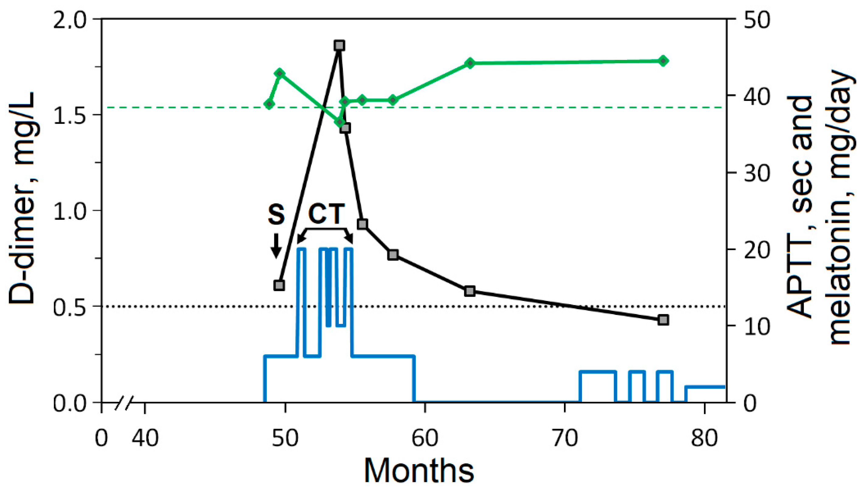
3.5.3. Hemostasis Disorders

Venous thromboembolism (VTE) is one of the most common causes of death and morbidity in cancer patients, and CT and immobilization contribute to an increased risk of VTE [76]. The prevalence of coagulopathy, including thrombocytopenia and APTT prolongation, has been shown in patients with CRC [6]. The elevated preoperative and postoperative level of D-dimer (the product of degradation of fibrin by fibrinolysis-mediated plasmin cleavage) and APTT may be risk factors associated with thrombotic events and bleeding, worsening the prognosis of CRC [77,78,79,80]. Cumulative incidences of VTE occur in 3.6% of CRC patients [81]. According to the Khorana scale, CRC is ranked as a cancer with a low thrombogenic risk among other types of organ-specific cancer; however, other authors believe that CRC has a higher thrombogenic risk [81,82]. The D-dimer test reflects the activation of fibrinolysis and thrombosis and is commonly used to evaluate patients with suspected deep venous thrombosis, pulmonary embolism, or disseminated intravascular coagulation. Testing of D-dimer in cancer patients has limitations since surgery, trauma, hemorrhage, infection, and other clinical conditions also increase its level [83]. Elevated levels of D-dimer are frequently observed in cancer patients; however, the test has lower specificity and a higher false-positive rate for VTE in patients compared to the general population. In addition, the clinical utility of D-dimer testing, as measured by the proportion of patients in whom VTE can be ruled out as a D-dimer-negative result, is much lower in cancer than in the non-cancer population. D-dimer levels increase with age, reducing the specificity of testing and requiring the use of an age-adjusted D-dimer cut-off. Thus, most cancer patients need additional diagnostic imaging to rule out VTE [83].
In the present study, the patient did not receive anticoagulant therapy, except for injections of low-molecular-weight heparin within 5 days after surgery for thromboprophylaxis [84]. The elevated postoperative level of D-dimer in the plasma increased during CT with Mel and then gradually decreased to the reference range (Figure 4). The APTT values tended to be above the upper limit in the follow-up. The plasma levels of fibrinogen and antithrombin III and the prothrombin time remained within the reference range. Ultrasonography of the patient did not find thrombotic events.
The influence of Mel on hemostasis and PLT is still poorly understood, and published data are contradictory. Administration of high doses of Mel can reduce thrombosis and levels of plasma fibrinogen but not D-dimer levels, as has been shown in patients infected with COVID-19 and in patients with hemorrhagic stroke [85,86]. In an experimental model, Mel was found to be able to shorten the APTT, prothrombin time, and thrombin time by modulating blood coagulation pathways [87]. Mel can increase the PLT count, including in patients receiving CT [88,89,90]. On the other hand, possible consequences of uncontrolled use of Mel have been reported since Mel can promote PLT aggregation and apoptosis [91]. A decrease in the number of PLTs was observed during CT in combination with Mel; therefore, CT was suspended (Figure 2). The patient was prescribed amoxicillin and azithromycin for the treatment of sinusitis and bronchitis. After treatment with antibiotics, a sharp but transient increase in the number of PLTs was observed (Figure 2, arrows Am-Az and Az). An increase in PLTs after taking antibiotics was noted three times: in the period between interrupted CT cycles and then after chemotherapy, including the period of Mel withdrawal. This is one of the side effects of antibiotics [92,93], which in the present case facilitated the prescription of subsequent CT cycles. The use of antibiotics to treat infections may increase the risk of venous thrombosis [94].
Antibiotics may be prescribed since infection is one of the concomitant diseases in cancer patients. Considering the essential role of the gut microbiota in the development and progression of colon cancer, antibiotics should be used with caution, as they can cause cancer-promoting dysbiosis and affect the efficacy and toxicity of CT by disturbing the balance and diversity of gut bacteria [95,96]. Both CT and antibiotics significantly affect the composition of intestinal bacteria, and bacteria can also influence the response to CT and IT [95,97]. Antibacterial activity against cancer-promoting Fusobacterium nucleatum has been found for 5-fluorouracil and aspirin, and individuals who use aspirin daily have a lower bacterial load in colon adenoma tissues [97,98]. This bacterium is an oral pathogen related to periodontal disease and can colonize and predominate in CRC tissue. This is associated with worse overall and cancer-specific survival [99,100]. Oral pathogenic bacteria may contribute to the progression of gastrointestinal cancer through the mechanisms of inflammation, tumor colonization, and dysregulation of the immune response [99]. In the present clinical case, the patient benefited from a publication on the effectiveness of Mel in maintaining oral health [101]. According to his own observations, periodontal inflammation was reduced after regular topical oral use of Mel.

4. Discussion

4.1. Relevance of the Use of Melatonin in Integrative Cancer Treatment

4.1.1. Physiological Function of Melatonin

The circadian system coordinates vital physiological functions and is synchronously connected with the visual perception of light and dark through the suprachiasmatic nucleus. Physiological functions of the body, including the normal functioning of the gastrointestinal tract, are maintained through circadian rhythms, the desynchronization of which can lead to gastrointestinal disorders and increase the risk of cancer [16,102,103]. Mel functions as a cellular protector and multifunctional hormone regulator that maintains homeostasis through vital circadian rhythms to protect the body from the development of different diseases [104]. Some authors consider Mel as the “cornerstone” on which neuroimmunoendocrinology has been built as an integral concept of homeostasis regulation [105]. Endogenous Mel is produced by the pineal gland mainly at night as well as by other organs, especially the gastrointestinal tract [106]. Mel regulates colonic motility, and the effect of exogenous Mel may be dose-dependent [106,107]. Its potential for the treatment of symptoms of irritable bowel syndrome and ulcerative colitis is being considered, although reports are conflicting. The treatment potential of Mel may be greater for gut disorders exacerbated by circadian disruption [107]. The physiological role of Mel and its therapeutic potential for gut diseases have been discussed in reviews [105,106,108].
Mel can potentially suppress cancer initiation, as a significant negative association between the use of Mel in older adults and the risk of CRC was documented in a population-based cohort study [109]. The disrupted sleep and circadian rhythms in cancer patients is closely associated with poor prognosis and treatment failure [110,111]. Since most cancer patients may suffer from various sleep disruptions, diagnosis and treatment of this comorbidity should not be ignored. Circadian modulation, sleep medication, and sleep-correcting interventions through a healthy daytime lifestyle should become an integrated practice in oncology [110]. Cortisol is a steroid hormone that is also considered a circadian hormone and is produced in response to stress. Cortisol is produced primarily in the adrenal cortex as well as in other tissues but in smaller quantities. Like Mel, cortisol is a multifunctional hormone regulator that maintains homeostasis. Circadian cortisol production may be disrupted in cancer, and the serum Mel/cortisol ratio may be reduced in patients with advanced cancer [110]. Poor sleep may lead to elevated serum cortisol levels and cortisol-mediated immunosuppression, but long-term disruption of circadian rhythms may deplete adrenal cortisol production. The effects of cortisol treatment and how it relates to circadian resynchronization remain unclear and deserve further research [110]. Distress and disruption of sleep and circadian rhythms contribute to tumor progression, as they deplete resources and cause disturbance of immune homeostasis, including conditions associated with dysbiosis and comorbidities [108]. The amplitude of the circadian rhythm in Mel secretion may be decreased in patients with CRC [112]. Regarding the effect on sleep and circadian rhythms, effective doses may be less than 2–3 mg/day, as administration of higher doses may cause the desensitization of Mel receptors [113,114].

4.1.2. Anticancer Activity of Melatonin and Its Preliminary Clinical Studies

The anticancer potential of Mel has been shown in many experimental works [16,17,115,116]. The mechanisms of antitumor activity of Mel are associated with pleiotropic effects on cancer cells and the tumor microenvironment, including the induction of apoptosis; anti-inflammatory, anti-angiogenic, antiproliferative, and antimetastatic action; and the regulation of the immune and psychoneuroendocrine systems [33,71,104,115,117,118,119,120,121,122,123,124,125,126,127,128].
Considering the information obtained in preclinical models, the doses and methods used are generally not adapted for clinical translation. In in vivo experiments, the doses of Mel generally exceed the doses acceptable for humans [129]. Due to poor solubility in aqueous media and the instability of Mel, the true concentrations of Mel tested in vitro can be significantly lower than those presented in some studies (1–10 mM) [129].
Preliminary clinical studies have shown that the use of Mel reduces the harmful effects of CT and RT [115,118,119,120,130,131,132]. In terms of protection against ionizing radiation, the use of Mel can be recommended in regular examinations using computed tomography [133]. According to some preliminary reports, Mel reduces the incidence of neurotoxicity, asthenia, thrombocytopenia, lymphocytopenia, and other side effects of CT [33,89,119,122] and may enhance the therapeutic effect of anticancer drugs [89,104,118,120,121,134]. If this is so, it would be possible to adjust needed doses of drugs or prescribe their continuous and longer cycles. In addition, the potential of Mel could be further explored for supportive and palliative therapy, at least to improve a patient’s quality of life [135].
In clinical studies by Lissoni et al., patients have taken oral Mel at high daily doses (≥20 mg) [70,71,88,89,118,119,120,121,125,136]. A favorable effect on the survival of patients with prostate cancer has been reported at a dose of 3 mg/day [137]. Thus, the question of optimal doses of Mel remains open. The oral route of administration may not be optimal because of lower bioavailability and individual variability [138,139]. Studies on relatively small groups of patients with terminal stages and different tumor localizations deserve attention and are provided in reviews [33,122]. In a study of 30 patients with metastatic CRC, the authors observed a higher percentage of disease control in the group of patients receiving irinotecan in combination with Mel [121]. The combined use of Mel with CT may increase the survival of patients with advanced non-small cell lung cancer (NSCLC) and gastrointestinal cancer [118,120,127,140]. Adjuvant Mel treatment has no effect on disease-free survival in patients with resected early-stage NSCLC but may confer prolonged survival in those with late-stage disease [141]. Notably, the positive effect of Mel on overall survival in prostate cancer patients has also been noted only in patients with an unfavorable prognosis [137]. In general, the benefit of treatment with Mel should be considered in individual cases, rather than in populations, with an assessment of the dynamics of tumor markers. For example, the oncostatic effect of Mel treatment may depend on the expression of the MT1 receptor subtype in the tumor, as has been shown in a case report involving hormone-refractory prostate cancer. Mel can affect the PSA level, which has been assessed as temporary stabilization of the disease [142].
The effect of Mel on immunocompetent cells and cytokine production in cancer patients remains poorly studied. An earlier preliminary study found differences in circulating cytokine levels, including decreased IL-6 levels, in patients with advanced solid tumors after 30 days of Mel administration (10 mg/day) [143]. Mel supplementation (20 mg/day) after 6 months did not affect NK cell cytotoxicity or phenotype, nor did it affect blood levels of inflammatory cytokines in patients with NSCLC [141]. The combination of IL-2 and Mel may stabilize the disease and increase the survival of patients with metastatic tumors [125,126,127,144]. The survival of patients with solid tumors and brain metastases who received RT of the brain was studied. Mel intake had no positive effect on survival compared with historical controls with a median of 4.1 months. The authors noted that the efficacy of Mel in advanced cancer is needed in further studies before a conclusion can be reached on the utility of Mel in this setting [145]. The effect of Mel on survival has not been found in patients with advanced NSCLC receiving CT. The authors note that studies are needed using a longer period of observation and a large sample size since their study included patients with a potentially poor prognosis with a median survival of 7.3 months [146].
The use of Mel supplements to improve sleep has increased significantly over the past decade in both healthy populations and cancer patients, which may be of interest for observational studies. The ability of Mel to improve sleep and quality of life has attracted attention as insomnia, anxiety, depression, pain, and other symptoms often accompany cancer. According to a meta-analysis, Mel might decrease chronic pain [147]. Bedtime Mel intake significantly reduced fatigue and the risk of depressive symptoms, improved quality of life and sleep, and increased expression of clock genes in women with breast cancer [148,149,150,151,152]. However, in patients with NSCLC who took Mel at a daily dose of 20 mg, no beneficial effects were observed in improving quality of life or reducing symptoms or side effects of CT and RT [141]. The beneficial effects of Mel users on quality of life and symptoms have not been confirmed by a systematic review and meta-analysis as well [153]. The authors believe that the main sources of heterogeneity leading to inconsistent results may be related to different types of cancer as well as methods and duration of treatment; therefore, large-scale randomized controlled trials including combinations, dosage, and duration of Mel administration deserve further study [153].
It should be taken into account that population-based studies are highly heterogeneous, and this poses a challenge for verifying the utility of integrative treatment. Stratification of the patients could reduce heterogeneity and inter-individual variability. However, the complex network of multifactorial interactions and abnormalities in cancer is a challenge despite large-scale analyses and can lead to inconsistent results. Currently, there is heterogeneity in the accumulated bioinformatics, which is due not only to inappropriate stratification, bias, quality of trials, or methods of assessment but also to the influence of numerous factors leading to different results in response to treatment. High heterogeneity may be due to tumor pathology itself, i.e., type, tumor localization, stages, gradation, genome instability and mutation, molecular and metabolic characteristics, and the type of treatment, doses, duration, bioavailability, and individual response to treatment associated with genetic polymorphism [154]. Moreover, concomitant diseases, the use of other drugs, physiological and psychosocial factors, and the composition of the resident microbiota may also affect the outcome of both CT and integrative treatment. In a critical multilateral analysis of reviews on the topic, “Melatonin and health,” the authors concluded that more systematic research is needed to understand and establish the connection between Mel and specific aspects of health [155]. Considering the safety and beneficial multifunctional effects of Mel as well as the decrease in its production with age [114,117,156,157,158], it is advisable to grant priority to clinical studies of Mel for the integrative treatment of cancer, in CT-mediated organ dysfunction, and in comorbid inflammatory and chronic diseases. Clinical studies of Mel and antioxidants in breast cancer are presented in a review [159]. Another patient-centered methodology, namely, the N-of-1 study design, is discussed in Section 4.3.

4.1.3. Safety and Bioavailability of Exogenous Melatonin

Mel intake is safe even in high doses, as mild to moderate adverse events have been recorded [104,160,161]. Studies with prespecified low risk of bias criteria for meta-analysis did not find an increase in serious adverse events or associated treatment discontinuation. Oral administration of Mel causes minor and short-term adverse events that can likely be avoided or controlled by reducing doses and time. The most frequently reported adverse events are related to fatigue, mood, or psychomotor and neurocognitive performance. A few studies have noted adverse events related to endocrine and cardiovascular function. The authors believe that future long-term trials and studies of its interactions with endogenous hormones and pharmaceuticals will confirm its safety [162,163].
The bioavailability of Mel varies and depends on its absorption, metabolism, and elimination. In oral administration, the bioavailability varies from 9 to 33% and is approximately 15%. A higher bioavailability of Mel has been recorded in its penetration through the mucous membranes of the nose and mouth [104,138,139]. The pharmacokinetics of rectal and other routes of administration of Mel have also been described [164]. Physiological concentrations of Mel in blood serum, measured at night in the elderly, are 0.10–0.51 nM. The use of Mel in pharmacological doses (0.4 and 4 mg) causes a multiple increase in its average maximal serum concentration to 1.75 and 17.24 nM, respectively, but Mel is rapidly eliminated, with a half-life of 45 min [138,165]. It should be noted that some herbal preparations can significantly inhibit the metabolism of Mel, increasing concentration and time of elimination [166]. Mel supplements may contain quantities other than those listed on the product label or may contain serotonin because, unlike medications, supplement production is not strictly controlled. In some countries, Mel is available only with a prescription and is considered a drug.

4.2. Integrative Approach to Cancer Treatment

Research in the field of cancer therapy is mainly focused on the eradication of cancer cells. Combined oncotherapy aimed at vulnerable tumor targets can be more efficacious compared to monotherapy [167]. The complete eradication of cancer cells is a challenge today because of their molecular heterogeneity, mutations, plasticity, and self-renewal and because of the resistance of cancer stem cells to CT. Metronomic CT, which is referred to as low-dose continuous CT administration with minimal drug interruption, can affect the phenotype of nonproliferating dormant cells through antiangiogenic, immunomodulatory, and other mechanisms [12,168]. Studies in this field need to provide evidence on the safety and efficacy of comparative trials and identifying suitable patients [169,170]. Some medications are worth attention as off-label drugs for repurposing to oncology since they may exert an influence on cancer-associated signaling and metabolic pathways through epigenetic mechanisms causing tumor dormancy (a long period of latency), differentiation, or communicative reprogramming [9,10,11,171,172]. Disseminated tumor cells can persist in a dormant state for years, or even decades, after treatment but remain a source of cancer recurrence. New strategies are being considered to prevent relapse by maintaining or eradicating dormant tumor cells [173]. Reprogramming of tumor systems (anakoinosis) using epigenetically active drugs to convert malignant tumor cells into a less aggressive state is considered a future biotherapeutic strategy for managing tumor resistance and inducing long-term remission [11,172]. The interaction of Mel with various types of stem cells is of increasing interest in terms of reprogramming cancer stem cells. The results in non-transformed stem cells cannot be generalized to cancer stem cells, as the observed effects of Mel are often opposite in non-tumor and tumor cells [174]. Mel regulates abnormal aerobic glycolysis, gluconeogenesis, the pentose phosphate pathway, and lipid metabolism inherent in the metabolic program of cancer cells as well as transcription factors (HIF-1α and p53) [175]. Thus, biological therapies create new opportunities to slow down further cancer progression by changing the pro-tumoral imbalance in the microenvironment of residual cancer cells after primary tumor resection. Biotherapy may be useful for patients who refuse or discontinue CT and RT due to adverse effects as well as for mitigating adverse effects.

4.2.1. Zinc and Selenium

The outcome of cancer patients and the effectiveness of therapy depend on the patient’s nutritional status [54]. Zinc and Se regulate numerous pathways in cancer and immune cells [176,177]. The mechanisms of the anticancer activity of zinc and its binding proteins are carried out through the stimulation of immune cells, the production of cytokines, the regulation of zinc-dependent enzymes, transporters and metallothioneins, apoptosis, transcription factors, gene expression, and signaling and through the influence on differentiation, proliferation, migration, inflammation, bioenergetic, metabolic, and other ways [176,178,179,180]. On the other hand, cancer cells need zinc to resist apoptosis through the mechanism of zinc-mediated caspase inhibition [176], and some zinc finger proteins (transcription factors) can both inhibit and promote cancer progression [180].
The bioavailability of zinc compounds depends on many factors, including gastrointestinal disturbances, inflammation, age, and type of diet (presence of absorption inhibitors in food) [181]. Zinc intake may improve liver function and reduce the risk of hepatocellular carcinoma, and its higher intake reduces the risk of colorectal and esophageal cancer [181,182]. The risk of mortality is significantly higher in laryngeal cancer patients with the lowest serum zinc levels compared with the highest [183]. Zinc deficiency has been found to be an independent predictor of gynecological cancer recurrence [184]. Experimental clinical studies of small groups showed a beneficial effect of zinc supplementation on the overall survival of patients with advanced nasopharyngeal carcinoma [185]. A critical review of pooled studies of zinc supplementation during cancer treatment noted a beneficial effect on mucositis after RT but not on CT-induced side effects. Overall, data from studies of larger cohorts rather argue against an effect of zinc on overall or disease-free survival, but the authors note heterogeneity in reported results [186]. Increased zinc intake during CT of patients with CRC may increase superoxide dismutase activity without affecting markers of oxidative stress [187]. An optimal intake of zinc could restore the normal immune response and reduce the risk of infection in immunocompromised cancer patients. However, the optimal immunostimulatory dose of zinc has not been determined. Moreover, an excess amount of zinc supplementation may cause immunosuppressive effects [176,178].
Se is an essential micronutrient, and its deficiency is harmful to human health [188]. The anticancer potential of Se is realized through various mechanisms, including redox balance and protection against ROS-mediated DNA damage, influence on gene expression, apoptosis, and cell cycle arrest, promotion of DNA repair, prevention of invasion and metastasis of tumor cells, and stimulation of the immune response [38,188]. Se compounds and their metabolites can act as antioxidants or pro-oxidants. Dietary organic Se, which is more bioavailable than its inorganic compounds, is incorporated into proteins to form selenoproteins, the latter functioning as oxidoreductases, synthetases, and Se transporters [38,189,190,191].
The anticancer activity of Se compounds and the synergistic effect with anticancer drugs observed in preclinical studies have prompted further clinical trials, alone or with standard therapy, to develop appropriate types, doses, and schedules [39,190,192,193,194,195,196,197,198]. Intake of Se (200 μg) in the form of Se yeast for 6 months may improve metabolic profiles and increase the percentage of regressed tumors, as has been shown in a small number of patients with cervical intraepithelial neoplasia [196].
Epidemiological studies have shown an increased incidence of cancer in populations with low Se intake and low plasma Se levels [193]. It has been reported that serum Se levels inversely relate to the risk of colorectal adenomas, severity of inflammatory bowel disease, and CRC [199]. The low level of Se in the serum of patients with colorectal and breast cancer is associated with advanced disease and a lower survival rate [40,200,201]. However, the chemopreventive effect of Se status on cancer risk and Se intake in clinical trials is still ambiguous and may be due to the heterogeneity of studies [38]. Cancer patients are often deficient in Se, and its supplementation is considered a promising option for radioprotection in RT without compromising the effectiveness of RT. No toxicity has been reported at doses of 0.3–0.5 mg/day, but it is recommended that Se status be determined in patients prior to RT [202,203]. High doses of sodium selenite can influence tumor chemoresistance and reduce the side effects of CT and RT, and tolerable doses of up to 5 mg have been studied in patients with advanced cancer [193]. In a Phase I study, doses up to 33 mg of sodium selenite as a single dose prior to RT were noted to be tolerable [204]. Some protocols have noted the safety and tolerability of high doses of sodium selenite, but the manifestation of its reversible toxicity still requires the selection of an appropriate dose and duration of treatment [193,194,197,202]. Naturally occurring organic Se compounds appear to have fewer side effects and fewer systemic effects compared to those reported for inorganic Se [177]. Methylselenocysteine and selenomethionine have been studied in clinical trials mainly in terms of their chemopreventive role in cancer [205]. Information on some Se compounds undergoing Phase I–III clinical trials is presented in review [190].
Se is a vital micronutrient for immune cells, and its deficiency impairs innate and adaptive immune responses. The benefits of Se supplementation for boosting immunity against pathogens, vaccination, or cancer in the general population have not yet received definitive support and need further comprehensive study, including redox mechanisms and the regulatory role of selenoproteins [191,206]. Due to a small safety window, excessive Se intake is still a subject of debate [188]. Increased intake of Se along with vitamins and minerals may be associated with an increased risk of mortality among patients with prostate cancer [207]. Thus, Se supplementation is beneficial for Se-deficient patients but requires proper monitoring of its levels since supranutritional doses might have adverse effects [208].

4.2.2. Vitamin D3

Assessment of vitamin D status is based on the determination of its metabolite 25-hydroxyvitamin D (calcitriol, 25(OH)D). Calcitriol, an active hormonal form of vitamin D, has been extensively studied in preclinical models and is well represented in a review [209]. Briefly, its anticancer activity is mediated by various signaling pathways, including the regulation of numerous genes via the vitamin D receptor, inhibition of proliferation, angiogenesis and metastases, induction of apoptosis, autophagy and differentiation, anti-inflammatory and antioxidant effects, and effects on cancer-associated stromal cells, cancer stem cells, and immune cells in the tumor microenvironment [209,210].
A chemopreventive effect of vitamin D intake and an inverse relationship between circulating 25(OH)D levels and the risk of colorectal adenoma and CRC have been reported in a large systematic review and meta-analysis [211]. CRC patients are often deficient in circulating 25(OH)D, and higher levels of 25(OH)D are associated with better survival [19,211,212,213,214]. Some authors do not support a chemopreventive effect of vitamin D supplementation in patients with colorectal neoplasms [215]. Despite encouraging experimental and epidemiological observations, discrepancies have been shown in the therapeutic effect of vitamin D supplementation and its influence on mortality in CRC patients [209,212]. Most reported observations are heterogeneous due to poor stratification of patients by cancer pathology. For example, preliminary results from the AMATERASU randomized trial suggest that vitamin D supplementation may improve survival in a subgroup of patients with p53-positive tumors of the digestive tract or poorly differentiated adenocarcinoma [216,217]. In addition, the effects of vitamin D may depend on the dose and individual differences such as bioavailability, vitamin D-related metabolism, vitamin D receptor genotype, and body mass index [209,210,218,219], which are usually overlooked and may be the cause of inconclusive results. Thus, the multifaceted effects of vitamin D and its dysregulated metabolism and function in cancer represent a challenge for population-based studies and require well-designed clinical trial schedules based on patient stratification, response to treatment at appropriate doses, monitoring of the vitamin D metabolites, and determination of the vitamin D receptor genotype.

4.2.3. Polyphenols

Plant-derived polyphenols are widely distributed and present in human food. The anticancer potential of polyphenols is well documented in preclinical models and has been examined for integrative treatment. Molecular mechanisms include modulation of signaling pathways, antioxidant and prooxidant activity, induction of apoptosis, autophagy, and cell cycle arrest; inhibition of proliferation of cancer cells, angiogenesis, invasiveness, and metastasis; and modulation of the immune response and anti-inflammatory effects [1,56,220,221,222,223]. The use of plant-derived polyphenols with anticancer drugs may reduce their toxicity and adverse effects or reverse the drug resistance of cancer cells [20,224]. However, caution should be exercised when using herbal products and CT drugs simultaneously. There is a risk of their interaction through the mechanisms of inhibition or induction of enzymes (the cytochrome P450 superfamily that metabolizes endogenous substrates and drugs) as well as through the effect on transport proteins (ATP-binding cassette drug transporters), which leads to a change in the pharmacokinetics of drugs [55,56,225]. Polyphenols could reduce intestinal and systemic inflammation through beneficial modification of the microbial community, which some authors consider a missing link in the mechanism of action of poorly bioavailable dietary polyphenols [56,226]. However, this concept requires detailed study since the action of polyphenols is not limited to pathogens [56].
The use of green tea extract after polypectomy may be beneficial for the prevention of colorectal adenoma [227,228]. Green tea consumption has been reported to reduce the risk of colorectal and other cancers, but evidence for its effect on cancer prevention and mortality has not been confirmed or has been limited to the highest category of green tea consumption compared to the lowest [45]. Despite promising results from preclinical studies, clinical studies have been inconclusive, which can be explained using high doses of polyphenols in preclinical models that appear to be beyond the reach of humans through diet and supplements [226]. In addition, clinical studies and meta-analysis of the association between plant polyphenol intake and the risk of developing CRC have revealed inconsistent results, which may be explained by low bioavailability, individual differences in the metabolism of polyphenols, their metabolic transformation depending on the composition of intestinal microbiota, and other factors [56,223,224,226,229,230]. For example, a nutrikinetic study of the oral administration of green tea polyphenols demonstrated interindividual variation in plasma concentrations, which could explain the partially contradictory effects observed in different populations or diseases [231].
Epigallocatechin gallate (EGCG) is one of the main green tea polyphenols, the anticancer activity of which has been confirmed in preclinical models [232]. Some clinical trials have shown that EGCG is generally well tolerated and may mitigate the harmful effects of RT [233]. Like other polyphenols, EGCG has poor bioavailability [232], but its use at high doses may evoke hepatotoxicity in rare cases. There are recommendations for users of green tea products to control liver function by ALT and AST [234]. The combination of EGCG with a high dose of Mel may alleviate or reduce the risk of possible EGCG hepatotoxicity [235]. In the present clinical case, the liver function of the patient was monitored by the markers ALT and AST, the values of which remained within the upper limits. Regarding an integrative approach to treatment, it is advisable to study such combinations that target vulnerable areas inherent in cancer and act through different mechanisms. Mel combined with some phytochemicals may have a complementary effect on tumor angiogenesis [13]. The enhanced oncostatic effect of Mel in combination with EGCG on cancer cells can be explained by the fact that the action of both occurs through different mechanisms [236].
Taxifolin (dihydroquercetin) is a polyphenol found in conifers (Siberian and Dahurian larch, French maritime pine, and others), in tamarind and milk thistle seeds, and in plant products (fruits, vegetables, oils, wines, etc.). The anticancer activity and mechanisms of taxifolin continue to be studied. Its dose-dependent ability to inhibit the viability, proliferation, migration, and invasion of cancer cells as well as to induce apoptosis through various mechanisms, including the Wnt/β-catenin signaling pathway, has been noted [237,238,239,240,241,242]. Taxifolin can prevent the development of obesity-induced hepatic steatosis, fibrogenesis, and tumorigenesis in a mouse model [243]. The protective efficacy of taxifolin against cisplatin-induced oxidative organ damage has been demonstrated in rodents [244,245,246]. In addition, taxifolin can inhibit P-glycoprotein-mediated transport activity in cancer cells, thereby reversing their multiresistance to chemotherapeutic agents. In this regard, taxifolin shows greater efficacy than green tea polyphenols and may increase the cytotoxicity of chemotherapeutic agents [247]. Taxifolin has a safety profile but low bioavailability [248]. Like other polyphenols, its concentrations and doses used in preclinical models have been significantly higher than those that could be achieved in clinical use.

4.2.4. Aspirin

The beneficial effect of aspirin in cancer can be explained by various mechanisms, including a reduction in metastatic spread and the incidence of venous thromboembolism (anti-inflammatory and antiplatelet-mediated action). These mechanisms are associated with the inhibition of cyclooxygenases COX-1 and COX-2, which are necessary for the synthesis of prostanoids [63,249,250,251]. Low doses of aspirin (75 mg/day) can prevent PLTs from binding to tumor cells, thereby inhibiting cancer cell proliferation and metastasis via PLT-derived signals [10]. COX-independent mechanisms, such as induction of apoptosis and blocking of the WNT-TCF pathway, are less studied [5,10], and discussion of this topic is beyond the scope of this review. According to observational studies, regular use of aspirin can reduce the risk of recurrent colorectal adenomas and cancer mortality, but the benefit of aspirin for cancer is still controversial [252,253,254,255]. The use of aspirin may increase the risk of gastrointestinal and cerebral bleeding; however, discontinuation of aspirin intake may increase the risk of vascular diseases [250]. Co-administration of aspirin with Mel may be beneficial in protecting the gastric mucosa from aspirin-induced damage [256].

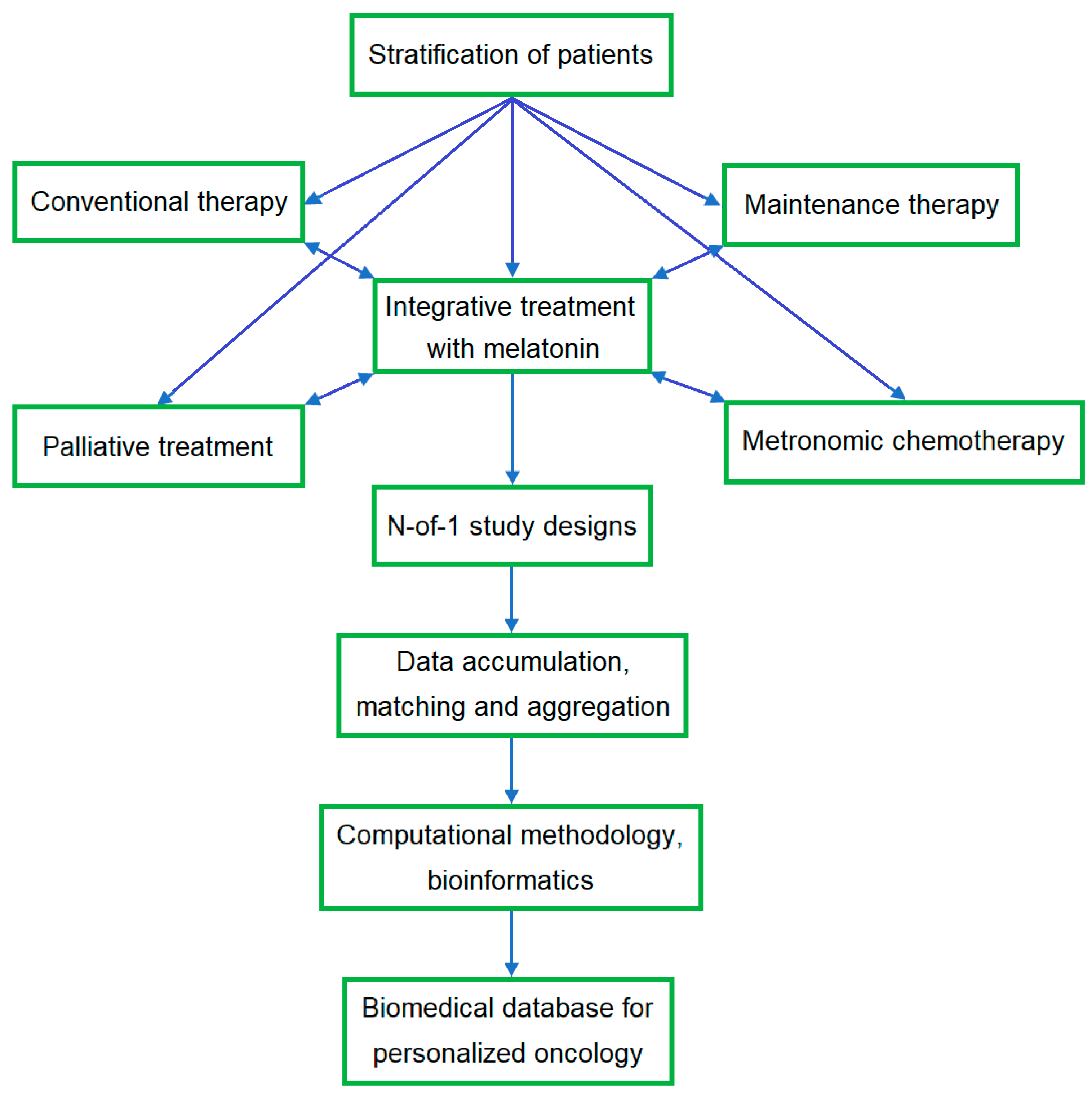
4.3. N-of-1 Study Design

The methodology of the N-of-1 trial represents a holistic approach to health accommodated to a patient-centered model rather than a drug-centered model [257,258]. The N-of-1 study design allows a treatment to be tailored to the patient depending on his response. Engaged patients can provide feedback and be more compliant towards the following recommendations. Recently, the N-of-1 trial design was upgraded to a high level of evidence for an individual’s treatment efficacy, placing this study design on par with a meta-analysis [259]. In terms of complementary treatment, N-of-1 studies are more suitable for treating symptoms of chronic and stable diseases [259]. N-of-1 trial designs can optimize treatment for patients, including those who cannot be included in RCTs due to uncertainty about the appropriate treatment pathway or for other reasons beyond the study criteria [259,260]. In N-of-1 studies, each participant serves as his or her own control. The duration of the N-of-1 study depends on the withdrawal period, i.e., washout to avoid carryover effects from the previous intervention and to properly assess the effect of the next intervention [260].
The power of N-of-1 studies depends on the number of clinical parameters measured rather than the number of participants and can help speed up cancer drug development, given its smaller number of participants and lower cost. One of the major limitations of N-of-1 studies is the inability to generalize the observed effects to the patient population. The effects of N-of-1 trials can be pooled and assessed in a stratified population, but this potential is considered a future prospect. Currently, N-of-1 studies remain heterogeneous, are useful as preliminary clinical data, and require a modified and standardized approach to estimate the efficacy of drugs used in oncology [257]. Dynamic studies and N-of-1 designs on individual patients have limitations for evidence-based medicine due to the influence of possible concomitant diseases requiring additional intervention. Repeated interventions using placebo and withdrawal periods demonstrating replication of the effect may reflect a causal relationship. Comparison and aggregation of serial studies of homogeneous groups with the same design may provide a more objective assessment and support for the hypothesized effect of integrative treatment.
Adequate personalized therapy involves studying the molecular genetic profile of the patient’s tumor and response to therapy. Evaluation of the Mel potential in integrative treatment using N-of-1 studies may involve a variety of designs, including its influence on CT effectiveness and adverse effects, tumor-associated systemic effects, and quality of life as well as tumor recurrence and survival (Figure 5). The N-of-1 study protocols may be considered for an RCT, similar to the trial evaluating the effectiveness of Mel in insomnia in children with neurological disorders [261]. Like Mel, natural supplements may also be examined using N-of-1 trials.

5. Conclusions

Numerous preclinical studies of adjunctive treatment of cancer using natural products have now been published, but these remain outside the scope of clinical practice. In clinical studies, special attention is drawn to non-toxic natural products that mitigate the harmful effects of CT and RT but do not affect the effectiveness of conventional therapy. Mel is considered a biomodulator that influences various pathways inherent in the initiation and progression of cancer. Preclinical and preliminary clinical studies suggest a dual effect of Mel in cancer treatment, as it may increase the effectiveness of anticancer drugs and mitigate their harmful effects on the body.
The present case report analyzes the dynamics and the well-being of a patient taking Mel for a long time, focusing on abnormal clinical parameters and adverse effects of CT. The use of Mel improved well-being and sleep, and its side effects were well tolerated. The patient’s condition during CT combined with Mel was satisfactory, and the polyneuropathy was temporary and manifested as a reaction to cold. The residual manifestation of polyneuropathy was a decrease in the sensitivity of the toes and soles. No effect of Mel supplements on thrombocytopenia was found, which may be associated with the patient’s predisposition to a lower PLT count. Inflammatory biomarkers (NLR, LMR, and PLR) varied after CT combined with Mel but remained within accepted favorable limits. The number of lymphocytes remained within the reference values and increased after surgery and during CT in combination with Mel. Postoperative levels of CEA and CA 19-9 were within the reference range. The PSA level decreased during CT in combination with Mel but increased after CT and especially to the end of Mel withdrawal and decreased after resuming Mel intake. The level of D-dimer increased during CT with Mel and then gradually decreased, while the APTT remained near the upper limits and then increased. A significant increase in the PLT count was observed as a reaction to treatment with azithromycin, including the Mel withdrawal period.
Regarding some recommendations and the results of preclinical and clinical studies, the patient took food supplements as well as aspirin after CT. Patients may use various combinations of antioxidants and nutritional supplements based on generally accepted information and their subjective perception and well-being. There are potential risks of simultaneous use of different medicines and herbal products, mainly related to their interactions, so the choice of treatment option should be made under the supervision of healthcare professionals and nutritionists [263].
Verification of the utility of Mel and natural supplements for integrative treatment in clinical settings is challenging due to the high heterogeneity and inter-individual variability of population-based studies. An alternative approach to a parallel group of RCTs may be N-of-1 study designs, with the potential to minimize costs and resources [264]. Multivariate and aggregated N-of-1 study designs are suitable for advancing biomedical and translational studies in precision medicine and can also provide participants with real-time care [260]. N-of-1 studies deserve attention to optimize the integrative treatment of complications caused by cancer and anticancer therapy and could be included in future medical research programs focused on collaboration with patient partners [265]. Retrospective and prospective studies of cancer patients treated with Mel in dynamic and N-of-1 designs based on protocols and questionnaires will help advance integrative treatment using Mel.

Author Contributions

Conceptualization, E.S.; methodology, E.S. and V.C.; software, E.S.; validation, E.S., V.C. and T.V.; investigation and analysis, E.S. and V.C.; resources, E.S., V.C. and T.V.; data curation, E.S. and V.C.; writing, review, and editing, E.S.; interpretation of data, E.S.; supervision, critical reviewing, and final approval of the version to be published, E.S., V.C. and T.V. All authors have read and agreed to the published version of the manuscript.

Funding

This research received no external funding.

Institutional Review Board Statement

The patient chose to remain outside the framework of institutional projects, respecting his confidentiality. The study was performed by the patient as a self-medication and self-management, with the aim of reducing the adverse effects of chemotherapy and improving well-being and quality of life, and was registered in the confidential protocols of the North Estonia Medical Centre as noted in the Materials and Methods (Section 2). Based on the results of the completed study, this paper was written taking into account the patient’s consent. The paper was approved by the Research Ethics Committee of the National Institute for Health Development in Estonia. NIHD REC, Confirmation No. 3/2024 dated 23 January 2024.

Informed Consent Statement

Informed consent for therapy was obtained from the patient involved in this study. The patient expressed his full consent to the analysis and publication of his clinical data, the results obtained, and the material of the present paper.

Data Availability Statement

The data are not publicly available for privacy reasons.

Acknowledgments

The authors are grateful to Vahur Valvere for helpful recommendations and Jaak Põder for consultations.

Conflicts of Interest

The authors declare no conflicts of interest.

References

  1. Bouyahya, A.; Mechchate, H.; Oumeslakht, L.; Zeouk, I.; Aboulaghras, S.; Balahbib, A.; Zengin, G.; Kamal, M.A.; Gallo, M.; Montesano, D.; et al. The Role of Epigenetic Modifications in Human Cancers and the Use of Natural Compounds as Epidrugs: Mechanistic Pathways and Pharmacodynamic Actions. Biomolecules 2022, 12, 367. [Google Scholar] [CrossRef] [PubMed]
  2. Kasprzak, A. The Role of Tumor Microenvironment Cells in Colorectal Cancer (CRC) Cachexia. Int. J. Mol. Sci. 2021, 22, 1565. [Google Scholar] [CrossRef] [PubMed]
  3. Lissoni, P.; Rovelli, F.; Vigorè, L.; Messina, G.; Lissoni, A.; Porro, G.; Di Fede, G. How to Monitor the Neuroimmune Biological Response in Patients Affected by Immune Alteration-Related Systemic Diseases. Methods Mol. Biol. 2018, 1781, 171–191. [Google Scholar] [CrossRef] [PubMed]
  4. Aguilar-Cazares, D.; Chavez-Dominguez, R.; Marroquin-Muciño, M.; Perez-Medina, M.; Benito-Lopez, J.J.; Camarena, A.; Rumbo-Nava, U.; Lopez-Gonzalez, J.S. The systemic-level repercussions of cancer-associated inflammation mediators produced in the tumor microenvironment. Front. Endocrinol. 2022, 13, 929572. [Google Scholar] [CrossRef] [PubMed]
  5. Zappavigna, S.; Cossu, A.M.; Grimaldi, A.; Bocchetti, M.; Ferraro, G.A.; Nicoletti, G.F.; Filosa, R.; Caraglia, M. Anti-Inflammatory Drugs as Anticancer Agents. Int. J. Mol. Sci. 2020, 21, 2605. [Google Scholar] [CrossRef] [PubMed]
  6. Admasu, F.T.; Dejenie, T.A.; Ayehu, G.W.; Zewde, E.A.; Dessie, G.; Adugna, D.G.; Enyew, E.F.; Geto, Z.; Abebe, E.C. Evaluation of thromboembolic event, basic coagulation parameters, and associated factors in patients with colorectal cancer: A multicenter study. Front. Oncol. 2023, 13, 1143122. [Google Scholar] [CrossRef] [PubMed]
  7. O’Brien, K.; Ried, K.; Binjemain, T.; Sali, A. Integrative Approaches to the Treatment of Cancer. Cancers 2022, 14, 5933. [Google Scholar] [CrossRef] [PubMed]
  8. Sung, H.; Ferlay, J.; Siegel, R.L.; Laversanne, M.; Soerjomataram, I.; Jemal, A.; Bray, F. Global Cancer Statistics 2020, GLOBOCAN Estimates of Incidence and Mortality Worldwide for 36 Cancers in 185 Countries. CA Cancer J. Clin. 2021, 71, 209–249. [Google Scholar] [CrossRef] [PubMed]
  9. Heudobler, D.; Lüke, F.; Vogelhuber, M.; Klobuch, S.; Pukrop, T.; Herr, W.; Gerner, C.; Pantziarka, P.; Ghibelli, L.; Reichle, A. Anakoinosis: Correcting Aberrant Homeostasis of Cancer Tissue-Going Beyond Apoptosis Induction. Front. Oncol. 2019, 9, 1408. [Google Scholar] [CrossRef] [PubMed]
  10. Nowak-Sliwinska, P.; Scapozza, L.; Ruiz i Altaba, A. Drug repurposing in oncology: Compounds, pathways, phenotypes and computational approaches for colorectal cancer. Biochim. Biophys. Acta Rev. Cancer 2019, 1871, 434–454. [Google Scholar] [CrossRef] [PubMed]
  11. Lüke, F.; Harrer, D.C.; Pantziarka, P.; Pukrop, T.; Ghibelli, L.; Gerner, C.; Reichle, A.; Heudobler, D. Drug Repurposing by Tumor Tissue Editing. Front. Oncol. 2022, 12, 900985. [Google Scholar] [CrossRef] [PubMed]
  12. Heudobler, D.; Rechenmacher, M.; Lüke, F.; Vogelhuber, M.; Klobuch, S.; Thomas, S.; Pukrop, T.; Hackl, C.; Herr, W.; Ghibelli, L.; et al. Clinical Efficacy of a Novel Therapeutic Principle, Anakoinosis. Front. Pharmacol. 2018, 9, 1357. [Google Scholar] [CrossRef] [PubMed]
  13. Block, K.I.; Gyllenhaal, C.; Lowe, L.; Amedei, A.; Amin, A.R.M.R.; Amin, A.; Aquilano, K.; Arbiser, J.; Arreola, A.; Arzumanyan, A. Designing a broad-spectrum integrative approach for cancer prevention and treatment. Semin. Cancer Biol. 2015, 35, S276–S304. [Google Scholar] [CrossRef]
  14. Bondy, S.C.; Campbell, A. Mechanisms Underlying Tumor Suppressive Properties of Melatonin. Int. J. Mol. Sci. 2018, 19, 2205. [Google Scholar] [CrossRef] [PubMed]
  15. Mihanfar, A.; Yousefi, B.; Azizzadeh, B.M. Interactions of melatonin with various signaling pathways: Implications for cancer therapy. Cancer Cell Int. 2022, 22, 420. [Google Scholar] [CrossRef]
  16. Chok, K.C.; Ng, C.H.; Koh, R.Y.; Ng, K.Y.; Chye, S.M. The potential therapeutic actions of melatonin in colorectal cancer. Horm. Mol. Biol. Clin. Investig. 2019, 39, 20190001. [Google Scholar] [CrossRef] [PubMed]
  17. Gil-Martín, E.; Egea, J.; Reiter, R.J.; Romero, A. The emergence of melatonin in oncology: Focus on colorectal cancer. Med. Res. Rev. 2019, 39, 2239–2285. [Google Scholar] [CrossRef]
  18. Evans, S.O.; Khairuddin, P.F.; Jameson, M.B. Optimising Selenium for Modulation of Cancer Treatments. Anticancer Res. 2017, 37, 6497–6509. [Google Scholar] [CrossRef] [PubMed]
  19. Maalmi, H.; Walter, V.; Jansen, L.; Boakye, D.; Schöttker, B.; Hoffmeister, M.; Brenner, H. Association between Blood 25-Hydroxyvitamin D Levels and Survival in Colorectal Cancer Patients: An Updated Systematic Review and Meta-Analysis. Nutrients 2018, 10, 896. [Google Scholar] [CrossRef]
  20. Arora, I.; Sharma, M.; Tollefsbol, T.O. Combinatorial Epigenetics Impact of Polyphenols and Phytochemicals in Cancer Prevention and Therapy. Int. J. Mol. Sci. 2019, 20, 4567. [Google Scholar] [CrossRef] [PubMed]
  21. Liu, S.; Lin, Y.; Huang, S.; Xue, S.; Huang, R.; Chen, L.; Wang, C. Identifying the long-term survival beneficiary of chemotherapy for stage N1c sigmoid colon cancer. Sci. Rep. 2022, 12, 16909. [Google Scholar] [CrossRef] [PubMed]
  22. Lin, Q.; Zhou, H.; Shi, S.; Lin, J.; Yan, W. The Prognostic Value of Adjuvant Chemotherapy in Colon Cancer with Solitary Tumor Deposit. Front. Oncol. 2022, 12, 916091. [Google Scholar] [CrossRef] [PubMed]
  23. Luo, Z.; Zhu, M.G.; Zhang, Z.Q.; Ye, F.J.; Huang, W.H.; Luo, X.Z. Increased expression of Ki-67 is a poor prognostic marker for colorectal cancer patients: A meta analysis. BMC Cancer 2019, 19, 123. [Google Scholar] [CrossRef] [PubMed]
  24. Xiong, D.-D.; Lin, X.-G.; He, R.-Q.; Pan, D.-H.; Luo, Y.-H.; Dang, Y.-W.; Luo, D.-Z.; Chen, G.; Peng, Z.-G.; Gan, T.-Q. Ki67/MIB-1 predicts better prognoses in colorectal cancer patients received both surgery and adjuvant radio-chemotherapy: A meta-analysis of 30 studies. Int. J. Clin. Exp. Med. 2017, 10, 1788–1804. [Google Scholar]
  25. Fluge, Ø.; Gravdal, K.; Carlsen, E.; Vonen, B.; Kjellevold, K.; Refsum, S.; Lilleng, R.; Eide, T.J.; Halvorsen, T.B.; Tveit, K.M.; et al. Norwegian Gastrointestinal Cancer Group. Expression of EZH2 and Ki-67 in colorectal cancer and associations with treatment response and prognosis. Br. J. Cancer 2009, 101, 1282–1289. [Google Scholar] [CrossRef] [PubMed]
  26. Munro, A.J.; Lain, S.; Lane, D.P. P53 abnormalities and outcomes in colorectal cancer: A systematic review. Br. J. Cancer 2005, 92, 434–444. [Google Scholar] [CrossRef] [PubMed]
  27. Williams, D.S.; Mouradov, D.; Browne, C.; Palmieri, M.; Elliott, M.J.; Nightingale, R.; Fang, C.G.; Li, R.; Mariadason, J.M.; Faragher, I. Overexpression of TP53 protein is associated with the lack of adjuvant chemotherapy benefit in patients with stage III colorectal cancer. Mod. Pathol. 2020, 33, 483–495. [Google Scholar] [CrossRef] [PubMed]
  28. Oh, H.J.; Bae, J.M.; Wen, X.; Jung, S.; Kim, Y.; Kim, K.J.; Cho, N.Y.; Kim, J.H.; Han, S.W.; Kim, T.Y. p53 expression status is associated with cancer-specific survival in stage III and high-risk stage II colorectal cancer patients treated with oxaliplatin-based adjuvant chemotherapy. Br. J. Cancer 2019, 120, 797–805. [Google Scholar] [CrossRef] [PubMed]
  29. Zaanan, A.; Cuilliere-Dartigues, P.; Guilloux, A.; Parc, Y.; Louvet, C.; de Gramont, A.; Tiret, E.; Dumont, S.; Gayet, B.; Validire, P. Impact of p53 expression and microsatellite instability on stage III colon cancer disease-free survival in patients treated by 5-fluorouracil and leucovorin with or without oxaliplatin. Ann. Oncol. 2010, 21, 772–780. [Google Scholar] [CrossRef] [PubMed]
  30. Ji, R.; Huang, G.; Xu, J.; Yu, X.; Zhou, A.; Du, C. Splenomegaly during oxaliplatin-based chemotherapy: Impact on blood parameters and anti-neoplastic treatment. Transl. Cancer Res. 2022, 11, 1880–1888. [Google Scholar] [CrossRef] [PubMed]
  31. Selvy, M.; Pereira, B.; Kerckhove, N.; Gonneau, C.; Feydel, G.; Pétorin, C.; Vimal-Baguet, A.; Melnikov, S.; Kullab, S.; Hebbar, M.; et al. Long-Term Prevalence of Sensory Chemotherapy-Induced Peripheral Neuropathy for 5 Years after Adjuvant FOLFOX Chemotherapy to Treat Colorectal Cancer: A Multicenter Cross-Sectional Study. J. Clin. Med. 2020, 9, 2400. [Google Scholar] [CrossRef] [PubMed]
  32. Areti, A.; Komirishetty, P.; Akuthota, M.; Malik, R.A.; Kumar, A. Melatonin prevents mitochondrial dysfunction and promotes neuroprotection by inducing autophagy during oxaliplatin-evoked peripheral neuropathy. J. Pineal. Res. 2017, 62, 3. [Google Scholar] [CrossRef]
  33. Wang, Y.; Wang, P.; Zheng, X.; Du, X. Therapeutic strategies of melatonin in cancer patients: A systematic review and meta-analysis. Onco Targets Ther. 2018, 11, 7895–7908. [Google Scholar] [CrossRef]
  34. Nahleh, Z.; Pruemer, J.; Lafollette, J.; Sweany, S. Melatonin, a promising role in taxane-related neuropathy. Clin. Med. Insights Oncol. 2010, 4, 35–41. [Google Scholar] [CrossRef]
  35. Ali, M.; Aziz, T. The Combination of Zinc and Melatonin Enhanced Neuroprotection and Attenuated Neuropathy in Oxaliplatin-Induced Neurotoxicity. Drug Des. Devel. Ther. 2022, 16, 3447–3463. [Google Scholar] [CrossRef] [PubMed]
  36. Dehmer, S.P.; O’Keefe, L.R.; Evans, C.V.; Guirguis-Blake, J.M.; Perdue, L.A.; Maciosek, M.V. Aspirin Use to Prevent Cardiovascular Disease and Colorectal Cancer: Updated Modeling Study for the US Preventive Services Task Force. JAMA. 2022, 327, 1598–1607. [Google Scholar] [CrossRef]
  37. Xiao, S.; Xie, W.; Fan, Y.; Zhou, L. Timing of Aspirin Use among Patients with Colorectal Cancer in Relation to Mortality: A Systematic Review and Meta-Analysis. JNCI Cancer Spectr. 2021, 5, pkab067. [Google Scholar] [CrossRef] [PubMed]
  38. Radomska, D.; Czarnomysy, R.; Radomski, D.; Bielawska, A.; Bielawski, K. Selenium as a Bioactive Micronutrient in the Human Diet and Its Cancer Chemopreventive Activity. Nutrients 2021, 13, 1649. [Google Scholar] [CrossRef] [PubMed]
  39. Hu, W.; Zhao, C.; Hu, H.; Yin, S. Food Sources of Selenium and Its Relationship with Chronic Diseases. Nutrients 2021, 13, 1739. [Google Scholar] [CrossRef] [PubMed]
  40. Baker, J.R.; Umesh, S.; Jenab, M.; Schomburg, L.; Tjønneland, A.; Olsen, A.; Boutron-Ruault, M.-C.; Rothwell, J.A.; Severi, G.; Katzke, V.; et al. Prediagnostic Blood Selenium Status and Mortality among Patients with Colorectal Cancer in Western European Populations. Biomedicines 2021, 9, 1521. [Google Scholar] [CrossRef] [PubMed]
  41. Pozharitskaya, O.N.; Karlina, M.V.; Shikov, A.N.; Kosman, V.M.; Makarova, M.N.; Makarov, V.G. Determination and pharmacokinetic study of taxifolin in rabbit plasma by high-performance liquid chromatography. Phytomedicine 2009, 16, 244–251. [Google Scholar] [CrossRef]
  42. Van Blarigan, E.L.; Meyerhardt, J.A. Role of physical activity and diet after colorectal cancer diagnosis. J. Clin. Oncol. 2015, 33, 1825–1834. [Google Scholar] [CrossRef] [PubMed]
  43. Fadelu, T.; Zhang, S.; Niedzwiecki, D.; Ye, X.; Saltz, L.B.; Mayer, R.J.; Mowat, R.B.; Whittom, R.; Hantel, A.; Benson, A.B.; et al. Nut Consumption and Survival in Patients with Stage III Colon Cancer: Results from CALGB 89803 (Alliance). J. Clin. Oncol. 2018, 36, 1112–1120. [Google Scholar] [CrossRef] [PubMed]
  44. Kristo, A.S.; Klimis-Zacas, D.; Sikalidis, A.K. Protective Role of Dietary Berries in Cancer. Antioxidants 2016, 5, 37. [Google Scholar] [CrossRef] [PubMed]
  45. Filippini, T.; Malavolti, M.; Borrelli, F.; Izzo, A.A.; Fairweather-Tait, S.J.; Horneber, M.; Vinceti, M. Green tea (Camellia sinensis) for the prevention of cancer. Cochrane Database Syst. Rev. 2020, 3, CD005004. [Google Scholar] [CrossRef]
  46. Mahomoodally, M.F.; Aumeeruddy, M.Z.; Rengasamy, K.R.R.; Roshan, S.; Hammad, S.; Pandohee, J.; Hu, X.; Zengin, G. Ginger and its active compounds in cancer therapy: From folk uses to nano-therapeutic applications. Semin. Cancer Biol. 2021, 69, 140–149. [Google Scholar] [CrossRef] [PubMed]
  47. Walter, V.; Jansen, L.; Ulrich, A.; Roth, W.; Bläker, H.; Chang-Claude, J.; Hoffmeister, M.; Brenner, H. Alcohol consumption and survival of colorectal cancer patients: A population-based study from Germany. Am. J. Clin. Nutr. 2016, 103, 1497–1506. [Google Scholar] [CrossRef] [PubMed]
  48. Romagnolo, D.F.; Donovan, M.G.; Doetschman, T.C.; Selmin, O.I. n-6 Linoleic Acid Induces Epigenetics Alterations Associated with Colonic Inflammation and Cancer. Nutrients 2019, 11, 171. [Google Scholar] [CrossRef] [PubMed]
  49. Nguyen, S.; Li, H.; Yu, D.; Cai, H.; Gao, J.; Gao, Y.; Luu, H.N.; Tran, H.; Xiang, Y.B.; Zheng, W. Dietary fatty acids and colorectal cancer risk in men: A report from the Shanghai Men’s Health Study and a meta-analysis. Int. J. Cancer 2021, 148, 77–89. [Google Scholar] [CrossRef] [PubMed]
  50. Simopoulos, A.P. The importance of the omega-6/omega-3 fatty acid ratio in cardiovascular disease and other chronic diseases. Exp. Biol. Med. 2008, 233, 674–688. [Google Scholar] [CrossRef]
  51. Bojková, B.; Winklewski, P.J.; Wszedybyl-Winklewska, M. Dietary Fat and Cancer-Which Is Good, Which Is Bad, and the Body of Evidence. Int. J. Mol. Sci. 2020, 21, 4114. [Google Scholar] [CrossRef] [PubMed]
  52. Shahjehan, F.; Merchea, A.; Cochuyt, J.J.; Li, Z.; Colibaseanu, D.T.; Kasi, P.M. Body Mass Index and Long-Term Outcomes in Patients with Colorectal Cancer. Front. Oncol. 2018, 8, 620. [Google Scholar] [CrossRef]
  53. Delpino, F.M.; Figueiredo, L.M. Melatonin supplementation and anthropometric indicators of obesity: A systematic review and meta-analysis. Nutrition 2021, 91–92, 111399. [Google Scholar] [CrossRef] [PubMed]
  54. Alam, W.; Ullah, H.; Santarcangelo, C.; Di Minno, A.; Khan, H.; Daglia, M.; Arciola, C.R. Micronutrient Food Supplements in Patients with Gastro-Intestinal and Hepatic Cancers. Int. J. Mol. Sci. 2021, 22, 8014. [Google Scholar] [CrossRef] [PubMed]
  55. Fasinu, P.S.; Rapp, G.K. Herbal Interaction with Chemotherapeutic Drugs-A Focus on Clinically Significant Findings. Front. Oncol. 2019, 9, 1356. [Google Scholar] [CrossRef] [PubMed]
  56. Duda-Chodak, A.; Tarko, T. Possible Side Effects of Polyphenols and Their Interactions with Medicines. Molecules 2023, 28, 2536. [Google Scholar] [CrossRef] [PubMed]
  57. Yasueda, A.; Urushima, H.; Ito, T. Efficacy and Interaction of Antioxidant Supplements as Adjuvant Therapy in Cancer Treatment: A Systematic Review. Integr. Cancer Ther. 2016, 15, 17–39. [Google Scholar] [CrossRef]
  58. Florido, J.; Rodriguez-Santana, C.; Martinez-Ruiz, L.; López-Rodríguez, A.; Acuña-Castroviejo, D.; Rusanova, I.; Escames, G. Understanding the Mechanism of Action of Melatonin, Which Induces ROS Production in Cancer Cells. Antioxidants 2022, 11, 1621. [Google Scholar] [CrossRef] [PubMed]
  59. Greten, F.R.; Grivennikov, S.I. Inflammation and Cancer: Triggers, Mechanisms, and Consequences. Immunity 2019, 51, 27–41. [Google Scholar] [CrossRef] [PubMed]
  60. Rumba, R.; Cipkina, S.; Cukure, F.; Vanags, A. Systemic and local inflammation in colorectal cancer. Acta Med. Litu. 2018, 25, 185–196. [Google Scholar] [CrossRef] [PubMed]
  61. Misiewicz, A.; Dymicka-Piekarska, V. Fashionable, but What is Their Real Clinical Usefulness? NLR, LMR, and PLR as a Propmising Indicator in Colorectal Cancer Prognosis: A Systematic Review. J. Inflamm. Res. 2023, 16, 69–81. [Google Scholar] [CrossRef] [PubMed]
  62. Yamamoto, T.; Kawada, K.; Obama, K. Inflammation-Related Biomarkers for the Prediction of Prognosis in Colorectal Cancer Patients. Int. J. Mol. Sci. 2021, 22, 8002. [Google Scholar] [CrossRef] [PubMed]
  63. Xu, X.R.; Yousef, G.M.; Ni, H. Cancer and platelet crosstalk: Opportunities and challenges for aspirin and other antiplatelet agents. Blood 2018, 131, 1777–1789. [Google Scholar] [CrossRef] [PubMed]
  64. Nielsen, S.R.; Schmid, M.C. Macrophages as Key Drivers of Cancer Progression and Metastasis. Mediators Inflamm. 2017, 2017, 9624760. [Google Scholar] [CrossRef] [PubMed]
  65. Liang, L.; Zhu, J.; Jia, H.; Huang, L.; Li, D.; Li, Q.; Li, X. Predictive value of pretreatment lymphocyte count in stage II colorectal cancer and in high-risk patients treated with adjuvant chemotherapy. Oncotarget 2016, 7, 1014–1028. [Google Scholar] [CrossRef] [PubMed]
  66. Yasui, K.; Shida, D.; Nakamura, Y.; Ahiko, Y.; Tsukamoto, S.; Kanemitsu, Y. Postoperative, but not preoperative, inflammation-based prognostic markers are prognostic factors in stage III colorectal cancer patients. Br. J. Cancer 2021, 124, 933–941. [Google Scholar] [CrossRef] [PubMed]
  67. Herold, Z.; Herold, M.; Lohinszky, J.; Szasz, A.M.; Dank, M.; Somogyi, A. Longitudinal changes in personalized platelet count metrics are good indicators of initial 3-year outcome in colorectal cancer. World J. Clin. Cases 2022, 10, 6825–6844. [Google Scholar] [CrossRef] [PubMed]
  68. Fu, J.; Zhu, J.; Du, F.; Zhang, L.; Li, D.; Huang, H.; Tian, T.; Liu, Y.; Zhang, L.; Liu, Y.; et al. Prognostic Inflammatory Index Based on Preoperative Peripheral Blood for Predicting the Prognosis of Colorectal Cancer Patients. Cancers 2021, 13, 3. [Google Scholar] [CrossRef] [PubMed]
  69. Li, Y.; Jia, H.; Yu, W.; Xu, Y.; Li, X.; Li, Q.; Cai, S. Nomograms for predicting prognostic value of inflammatory biomarkers in colorectal cancer patients after radical resection. Int. J. Cancer 2016, 139, 220–231. [Google Scholar] [CrossRef] [PubMed]
  70. Lissoni, P.; Messina, G.; Borsotti, G.; Alessio, T.; Stefano, F.; Simonetta, T.; Di Fede, G. Modulation of Immune and Anti-Tumor Effects of Cancer Immunotherapy with Anti-Pd-1 Monoclonal Antibodies by the Pineal Hormone Melatonin: Preliminary Clinical Results. J. Immuno. Allerg. 2020, 1, 1–6. [Google Scholar] [CrossRef]
  71. Lissoni, P.; Barni, S.; Tancini, G.; Ardizzoia, A.; Ricci, G.; Aldeghi, R.; Brivio, F.; Tisi, E.; Rovelli, F.; Rescaldani, R.; et al. A randomised study with subcutaneous low-dose interleukin 2 alone vs interleukin 2 plus the pineal neurohormone melatonin in advanced solid neoplasms other than renal cancer and melanoma. Br. J. Cancer 1994, 69, 196–199. [Google Scholar] [CrossRef] [PubMed]
  72. Noh, O.K.; Oh, S.Y.; Kim, Y.B.; Suh, K.W. Prognostic Significance of Lymphocyte Counts in Colon Cancer Patients Treated with FOLFOX Chemotherapy. World J. Surg. 2017, 41, 2898–2905. [Google Scholar] [CrossRef] [PubMed]
  73. Idos, G.E.; Kwok, J.; Bonthala, N.; Kysh, L.; Gruber, S.B.; Qu, C. The Prognostic Implications of Tumor Infiltrating Lymphocytes in Colorectal Cancer: A Systematic Review and Meta-Analysis. Sci. Rep. 2020, 10, 3360. [Google Scholar] [CrossRef] [PubMed]
  74. Wei, Y.; Zhang, X.; Wang, G.; Zhou, Y.; Luo, M.; Wang, S.; Hong, C. The impacts of pretreatment circulating eosinophils and basophils on prognosis of stage I-III colorectal cancer. Asia. Pac. J. Clin. Oncol. 2018, 14, e243–e251. [Google Scholar] [CrossRef] [PubMed]
  75. Gao, L.; Yuan, C.; Fu, J.; Tian, T.; Huang, H.; Zhang, L.; Li, D.; Liu, Y.; Meng, S.; Liu, Y. Prognostic scoring system based on eosinophil- and basophil-related markers for predicting the prognosis of patients with stage II and stage III colorectal cancer: A retrospective cohort study. Front. Oncol. 2023, 13, 1182944. [Google Scholar] [CrossRef] [PubMed]
  76. Akinbo, D.B.; Ajayi, O.I. Thrombotic Pathogenesis and Laboratory Diagnosis in Cancer Patients, An Update. Int. J. Gen. Med. 2023, 16, 259–272. [Google Scholar] [CrossRef] [PubMed]
  77. Lu, S.L.; Ye, Z.H.; Ling, T.; Liang, S.Y.; Li, H.; Tang, X.Z.; Xu, Y.S.; Tang, W.Z. High pretreatment plasma D-dimer predicts poor survival of colorectal cancer: Insight from a meta-analysis of observational studies. Oncotarget 2017, 8, 81186–81194. [Google Scholar] [CrossRef] [PubMed]
  78. Liu, C.; Ning, Y.; Chen, X.; Zhu, Q. D-Dimer level was associated with prognosis in metastatic colorectal cancer: A Chinese patients based cohort study. Medicine 2020, 99, e19243. [Google Scholar] [CrossRef] [PubMed]
  79. Shibutani, M.; Kashiwagi, S.; Fukuoka, T.; Iseki, Y.; Kasashima, H.; Kitayama, K.; Maeda, K. Prognostic Role of Preoperative D-dimer Levels in Patients with Stage I-III Colorectal Cancer. Cancer Diagn. Progn. 2023, 3, 38–43. [Google Scholar] [CrossRef] [PubMed]
  80. Zhang, L.; Ye, J.; Luo, Q.; Kuang, M.; Mao, M.; Dai, S.; Wang, X. Prediction of Poor Outcomes in Patients with Colorectal Cancer: Elevated Preoperative Prothrombin Time (PT) and Activated Partial Thromboplastin Time (APTT). Cancer Manag. Res. 2020, 12, 5373–5384. [Google Scholar] [CrossRef]
  81. Tønnesen, J.; Pallisgaard, J.; Rasmussen, P.V.; Ruwald, M.H.; Lamberts, M.; Nouhravesh, N.; Strange, J.; Gislason, G.H.; Hansen, M.L. Risk and timing of venous thromboembolism in patients with gastrointestinal cancer: A nationwide Danish cohort study. BMJ Open 2023, 13, e062768. [Google Scholar] [CrossRef] [PubMed]
  82. Khorana, A.A.; Kuderer, N.M.; Culakova, E.; Lyman, G.H.; Francis, C.W. Development and validation of a predictive model for chemotherapy-associated thrombosis. Blood 2008, 111, 4902–4907. [Google Scholar] [CrossRef] [PubMed]
  83. Peterson, E.A.; Lee, A.Y.Y. Update from the clinic: What’s new in the diagnosis of cancer-associated thrombosis? Hematology Am. Soc. Hematol. Educ. Program. 2019, 2019, 167–174. [Google Scholar] [CrossRef] [PubMed]
  84. Bosch, F.T.M.; Mulder, F.I.; Kamphuisen, P.W.; Middeldorp, S.; Bossuyt, P.M.; Büller, H.R.; van Es, N. Primary thromboprophylaxis in ambulatory cancer patients with a high Khorana score: A systematic review and meta-analysis. Blood Adv. 2020, 4, 5215–5225. [Google Scholar] [CrossRef] [PubMed]
  85. Hasan, Z.T.; Atrakji, D.M.Q.Y.M.A.A.; Mehuaiden, D.A.K. The Effect of Melatonin on Thrombosis, Sepsis and Mortality Rate in COVID-19 Patients. Int. J. Infect. Dis. 2022, 114, 79–84. [Google Scholar] [CrossRef] [PubMed]
  86. Shohrati, M.; Mohammadi, A.; Najafi, A.; Sharifzadeh, M.; Sharifnia, H.; Abdollahi, M.; Salesi, M.; Sahebnasagh, A.; Mojtahedzadeh, M. Evaluation of the Effects of Melatonin Supplementation on Coagulation in Patients with Haemorrhagic Stroke; A Randomized, Double-Blind, Controlled Trial. Front. Emerg. Med. 2021, 5, e32. [Google Scholar] [CrossRef]
  87. Pashalieva, I.; Decheva, L.; Stancheva, E.; Nyagolov, Y.; Negrev, N. Melatonin and luzindole–induced effects on integral blood coagulation parameters in rats. Comptes Rendus Acad. Bulg. Sci. 2014, 67, 1269–1274. [Google Scholar]
  88. Lissoni, P.; Mandala, M.; Rossini, F.; Fumagalli, L.; Barni, S. Growth Factors: Thrombopoietic Property of the Pineal Hormone Melatonin. Hematology 1999, 4, 335–343. [Google Scholar] [CrossRef] [PubMed]
  89. Lissoni, P.; Barni, S.; Mandalà, M.; Ardizzoia, A.; Paolorossi, F.; Vaghi, M.; Longarini, R.; Malugani, F.; Tancini, G. Decreased toxicity and increased efficacy of cancer chemotherapy using the pineal hormone melatonin in metastatic solid tumour patients with poor clinical status. Eur. J. Cancer 1999, 35, 1688–1692. [Google Scholar] [CrossRef]
  90. Esmaeili, A.; Nassiri Toosi, M.; Taher, M.; Bayani, J.; Namazi, S. Melatonin effect on platelet count in patients with liver disease. Gastroenterol. Hepatol. Bed Bench 2021, 14, 356–361. [Google Scholar] [PubMed]
  91. Girish, K.S.; Paul, M.; Thushara, R.M.; Hemshekhar, M.; Shanmuga Sundaram, M.; Rangappa, K.S.; Kemparaju, K. Melatonin elevates apoptosis in human platelets via ROS mediated mitochondrial damage. Biochem. Biophys. Res. Commun. 2013, 438, 198–204. [Google Scholar] [CrossRef] [PubMed]
  92. Frenck, R.W., Jr.; Mansour, A.; Nakhla, I.; Sultan, Y.; Putnam, S.; Wierzba, T.; Morsy, M.; Knirsch, C. Short-course azithromycin for the treatment of uncomplicated typhoid fever in children and adolescents. Clin. Infect. Dis. 2004, 38, 951–957. [Google Scholar] [CrossRef] [PubMed]
  93. Yang, C.J.; Hwang, J.J.; Hung, J.Y.; Chong, I.W.; Huang, M.S. Extreme thrombocytosis under the treatment by amoxicillin/clavulanate. Pharm. World Sci. 2006, 28, 326–328. [Google Scholar] [CrossRef] [PubMed]
  94. Timp, J.F.; Cannegieter, S.C.; Tichelaar, V.; Braekkan, S.K.; Rosendaal, F.R.; le Cessie, S.; Lijfering, W.M. Antibiotic use as a marker of acute infection and risk of first and recurrent venous thrombosis. Br. J. Haematol. 2017, 176, 961–970. [Google Scholar] [CrossRef] [PubMed]
  95. Panebianco, C.; Andriulli, A.; Pazienza, V. Pharmacomicrobiomics: Exploiting the drug-microbiota interactions in anticancer therapies. Microbiome 2018, 6, 92. [Google Scholar] [CrossRef] [PubMed]
  96. Mohamed, A.; Menon, H.; Chulkina, M.; Yee, N.S.; Pinchuk, I.V. Drug–Microbiota Interaction in Colon Cancer Therapy: Impact of Antibiotics. Biomedicines 2021, 9, 259. [Google Scholar] [CrossRef] [PubMed]
  97. LaCourse, K.D.; Zepeda-Rivera, M.; Kempchinsky, A.G.; Baryiames, A.; Minot, S.S.; Johnston, C.D.; Bullman, S. The cancer chemotherapeutic 5-fluorouracil is a potent Fusobacterium nucleatum inhibitor and its activity is modified by intratumoral microbiota. Cell Rep. 2022, 41, 111625. [Google Scholar] [CrossRef]
  98. Brennan, C.A.; Nakatsu, G.; Gallini Comeau, C.A.; Drew, D.A.; Glickman, J.N.; Schoen, R.E.; Chan, A.T.; Garrett, W.S. Aspirin Modulation of the Colorectal Cancer-Associated Microbe Fusobacterium nucleatum. mBio 2021, 12, e00547-21. [Google Scholar] [CrossRef] [PubMed]
  99. Smorodin, E.P. Prospects and Challenges of the Study of Anti-Glycan Antibodies and Microbiota for the Monitoring of Gastrointestinal Cancer. Int. J. Mol. Sci. 2021, 22, 11608. [Google Scholar] [CrossRef] [PubMed]
  100. Huangfu, S.C.; Zhang, W.B.; Zhang, H.R.; Li, Y.; Zhang, Y.R.; Nie, J.L.; Chu, X.; Chen, C.S.; Jiang, H.P.; Pan, J.H. Clinicopathological and prognostic significance of Fusobacterium nucleatum infection in colorectal cancer: A meta-analysis. J. Cancer 2021, 12, 1583–1591. [Google Scholar] [CrossRef]
  101. Liu, R.Y.; Li, L.; Zhang, Z.T.; Wu, T.; Lin, S.; Zhang, X.T. Clinical efficacy of melatonin as adjunctive therapy to non-surgical treatment of periodontitis: A systematic review and meta-analysis. Inflammopharmacology 2022, 30, 695–704. [Google Scholar] [CrossRef] [PubMed]
  102. Fowler, S.; Hoedt, E.C.; Talley, N.J.; Keely, S.; Burns, G.L. Circadian Rhythms and Melatonin Metabolism in Patients with Disorders of Gut-Brain Interactions. Front. Neurosci. 2022, 16, 825246. [Google Scholar] [CrossRef]
  103. Liu, W.; Zhou, Z.; Dong, D.; Sun, L.; Zhang, G. Sex Differences in the Association between Night Shift Work and the Risk of Cancers: A Meta-Analysis of 57 Articles. Dis. Markers 2018, 2018, 7925219. [Google Scholar] [CrossRef] [PubMed]
  104. Talib, W.H.; Alsayed, A.R.; Abuawad, A.; Daoud, S.; Mahmod, A.I. Melatonin in Cancer Treatment: Current Knowledge and Future Opportunities. Molecules 2021, 26, 2506. [Google Scholar] [CrossRef] [PubMed]
  105. Kvetnoy, I.; Ivanov, D.; Mironova, E.; Evsyukova, I.; Nasyrov, R.; Kvetnaia, T.; Polyakova, V. Melatonin as the Cornerstone of Neuroimmunoendocrinology. Int. J. Mol. Sci. 2022, 23, 1835. [Google Scholar] [CrossRef] [PubMed]
  106. Esteban-Zubero, E.; López-Pingarrón, L.; Alatorre-Jiménez, M.A.; Ochoa-Moneo, P.; Buisac-Ramón, C.; Rivas-Jiménez, M.; Castán-Ruiz, S.; Antoñanzas-Lombarte, Á.; Tan, D.X.; García, J.J.; et al. Melatonin’s role as a co-adjuvant treatment in colonic diseases: A review. Life Sci. 2017, 170, 72–81. [Google Scholar] [CrossRef] [PubMed]
  107. Hibberd, T.J.; Ramsay, S.; Spencer-Merris, P.; Dinning, P.G.; Zagorodnyuk, V.P.; Spencer, N.J. Circadian rhythms in colonic function. Front. Physiol. 2023, 14, 1239278. [Google Scholar] [CrossRef] [PubMed]
  108. Iesanu, M.I.; Zahiu, C.D.M.; Dogaru, I.-A.; Chitimus, D.M.; Pircalabioru, G.G.; Voiculescu, S.E.; Isac, S.; Galos, F.; Pavel, B.; O’Mahony, S.M.; et al. Melatonin–Microbiome Two-Sided Interaction in Dysbiosis-Associated Conditions. Antioxidants 2022, 11, 2244. [Google Scholar] [CrossRef] [PubMed]
  109. Zhang, N.; Sundquist, J.; Sundquist, K.; Ji, J. Use of Melatonin Is Associated with Lower Risk of Colorectal Cancer in Older Adults. Clin. Transl. Gastroenterol. 2021, 12, e00396. [Google Scholar] [CrossRef] [PubMed]
  110. Jensen, L.D.; Oliva, D.; Andersson, B.Å.; Lewin, F. A multidisciplinary perspective on the complex interactions between sleep, circadian, and metabolic disruption in cancer patients. Cancer Metastasis Rev. 2021, 40, 1055–1071. [Google Scholar] [CrossRef] [PubMed]
  111. Innominato, P.F.; Giacchetti, S.; Bjarnason, G.A.; Focan, C.; Garufi, C.; Coudert, B.; Iacobelli, S.; Tampellini, M.; Durando, X.; Mormont, M.C.; et al. Prediction of overall survival through circadian rest-activity monitoring during chemotherapy for metastatic colorectal cancer. Int. J. Cancer 2012, 131, 2684–2692. [Google Scholar] [CrossRef] [PubMed]
  112. Kos-Kudla, B.; Ostrowska, Z.; Kozlowski, A.; Marek, B.; Ciesielska-Kopacz, N.; Kudla, M.; Kajdaniuk, D.; Strzelczyk, J.; Staszewicz, P. Circadian rhythm of melatonin in patients with colorectal carcinoma. Neuro. Endocrinol. Lett. 2002, 23, 239–242. [Google Scholar] [PubMed]
  113. Minich, D.M.; Henning, M.; Darley, C.; Fahoum, M.; Schuler, C.B.; Frame, J. Is Melatonin the “Next Vitamin D”?: A Review of Emerging Science, Clinical Uses, Safety, and Dietary Supplements. Nutrients 2022, 14, 3934. [Google Scholar] [CrossRef] [PubMed]
  114. Anghel, L.; Baroiu, L.; Popazu, C.R.; Pătraș, D.; Fotea, S.; Nechifor, A.; Ciubara, A.; Nechita, L.; Mușat, C.L.; Stefanopol, I.A.; et al. Benefits and adverse events of melatonin use in the elderly (Review). Exp. Ther. Med. 2022, 23, 219. [Google Scholar] [CrossRef] [PubMed]
  115. Gurunathan, S.; Qasim, M.; Kang, M.H.; Kim, J.H. Role and Therapeutic Potential of Melatonin in Various Type of Cancers. Onco Targets Ther. 2021, 14, 2019–2052. [Google Scholar] [CrossRef] [PubMed]
  116. Xin, Z.; Jiang, S.; Jiang, P.; Yan, X.; Fan, C.; Di, S.; Wu, G.; Yang, Y.; Reiter, R.J.; Ji, G. Melatonin as a treatment for gastrointestinal cancer: A review. J. Pineal Res. 2015, 58, 375–387. [Google Scholar] [CrossRef] [PubMed]
  117. Zarezadeh, M.; Khorshidi, M.; Emami, M.; Janmohammadi, P.; Kord-Varkaneh, H.; Mousavi, S.M.; Mohammed, S.H.; Saedisomeolia, A.; Alizadeh, S. Melatonin supplementation and pro-inflammatory mediators: A systematic review and meta-analysis of clinical trials. Eur. J. Nutr. 2020, 59, 1803–1813. [Google Scholar] [CrossRef] [PubMed]
  118. Lissoni, P. Biochemotherapy with standard chemotherapies plus the pineal hormone melatonin in the treatment of advanced solid neoplasms. Pathol. Biol. 2007, 55, 201–204. [Google Scholar] [CrossRef] [PubMed]
  119. Lissoni, P. Is there a role for melatonin in supportive care? Support. Care Cancer 2002, 10, 110–116. [Google Scholar] [CrossRef] [PubMed]
  120. Lissoni, P.; Chilelli, M.; Villa, S.; Cerizza, L.; Tancini, G. Five years survival in metastatic non-small cell lung cancer patients treated with chemotherapy alone or chemotherapy and melatonin: A randomized trial. J. Pineal Res. 2003, 35, 12–15. [Google Scholar] [CrossRef]
  121. Cerea, G.; Vaghi, M.; Ardizzoia, A.; Villa, S.; Bucovec, R.; Mengo, S.; Gardani, G.; Tancini, G.; Lissoni, P. Biomodulation of cancer chemotherapy for metastatic colorectal cancer: A randomized study of weekly low-dose irinotecan alone versus irinotecan plus the oncostatic pineal hormone melatonin in metastatic colorectal cancer patients progressing on 5-fluorouracil-containing combinations. Anticancer Res. 2003, 23, 1951–1954. [Google Scholar] [PubMed]
  122. Seely, D.; Wu, P.; Fritz, H.; Kennedy, D.A.; Tsui, T.; Seely, A.J.; Mills, E. Melatonin as adjuvant cancer care with and without chemotherapy: A systematic review and meta-analysis of randomized trials. Integr. Cancer Ther. 2012, 11, 293–303. [Google Scholar] [CrossRef] [PubMed]
  123. Wu, H.; Liu, J.; Yin, Y.; Zhang, D.; Xia, P.; Zhu, G. Therapeutic Opportunities in Colorectal Cancer: Focus on Melatonin Antioncogenic Action. Biomed. Res. Int. 2019, 2019, 9740568. [Google Scholar] [CrossRef] [PubMed]
  124. Su, S.C.; Hsieh, M.J.; Yang, W.E.; Chung, W.H.; Reiter, R.J.; Yang, S.F. Cancer metastasis: Mechanisms of inhibition by melatonin. J. Pineal Res. 2017, 62, 1. [Google Scholar] [CrossRef]
  125. Lissoni, P.; Rovelli, F.; Porro, G.; Brivio, F.; Fumagalli, L.; Lissoni, A.; Messina, G.; Cenaj, V.; Di Fede, G. Treatment of Advanced Cancer related Lymphocytopenia: Comparison among the Effects of Subcutaneous Low-dose Interleukin-2, High-dose Pineal Hormone Melatonin and Checkpoint Inhibitors. J. Cancer Res. Oncobiol. 2018, 1, 112. [Google Scholar] [CrossRef]
  126. Vigoré, L.; Messina, G.; Brivio, F.; Fumagalli, L.; Rovelli, F.; Di Fede, G.; Lissoni, P. Psychoneuroendocrine modulation of regulatory T lymphocyte system: In vivo and in vitro effects of the pineal immunomodulating hormone melatonin. In Vivo 2010, 24, 787–789. [Google Scholar] [PubMed]
  127. Lissoni, P.; Messina, G.; Lissoni, A.; Franco, R. The psychoneuroendocrine-immunotherapy of cancer: Historical evolution and clinical results. J. Res. Med. Sci. 2017, 22, 45. [Google Scholar] [CrossRef] [PubMed]
  128. Bitzer-Quintero, O.K.; Ortiz, G.G.; Jaramillo-Bueno, S.; Ramos-González, E.J.; Márquez-Rosales, M.G.; Delgado-Lara, D.L.C.; Torres-Sánchez, E.D.; Tejeda-Martínez, A.R.; Ramirez-Jirano, J. Psycho-Neuro-Endocrine-Immunology: A Role for Melatonin in This New Paradigm. Molecules 2022, 27, 4888. [Google Scholar] [CrossRef] [PubMed]
  129. Li, Y.; Li, S.; Zhou, Y.; Meng, X.; Zhang, J.J.; Xu, D.P.; Li, H.B. Melatonin for the prevention and treatment of cancer. Oncotarget 2017, 8, 39896–39921. [Google Scholar] [CrossRef] [PubMed]
  130. Ma, Z.; Xu, L.; Liu, D.; Zhang, X.; Di, S.; Li, W.; Zhang, J.; Reiter, R.J.; Han, J.; Li, X.; et al. Utilizing Melatonin to Alleviate Side Effects of Chemotherapy: A Potentially Good Partner for Treating Cancer with Ageing. Oxid. Med. Cell. Longev. 2020, 2020, 6841581. [Google Scholar] [CrossRef] [PubMed]
  131. Onseng, K.; Johns, N.P.; Khuayjarernpanishk, T.; Subongkot, S.; Priprem, A.; Hurst, C.; Johns, J. Beneficial Effects of Adjuvant Melatonin in Minimizing Oral Mucositis Complications in Head and Neck Cancer Patients Receiving Concurrent Chemoradiation. J. Altern. Complement. Med. 2017, 23, 957–963. [Google Scholar] [CrossRef] [PubMed]
  132. Farhood, B.; Goradel, N.H.; Mortezaee, K.; Khanlarkhani, N.; Salehi, E.; Nashtaei, M.S.; Mirtavoos-Mahyari, H.; Motevaseli, E.; Shabeeb, D.; Musa, A.E.; et al. Melatonin as an adjuvant in radiotherapy for radioprotection and radiosensitization. Clin. Transl. Oncol. 2019, 21, 268–279. [Google Scholar] [CrossRef] [PubMed]
  133. Nuszkiewicz, J.; Woźniak, A.; Szewczyk-Golec, K. Ionizing Radiation as a Source of Oxidative Stress—The Protective Role of Melatonin and Vitamin D. Int. J. Mol. Sci. 2020, 21, 5804. [Google Scholar] [CrossRef] [PubMed]
  134. Jadid, M.F.S.; Aghaei, E.; Taheri, E.; Seyyedsani, N.; Chavoshi, R.; Abbasi, S.; Khorrami, A.; Goleij, P.; Hajazimian, S.; Taefehshokr, S.; et al. Melatonin increases the anticancer potential of doxorubicin in Caco-2 colorectal cancer cells. Environ. Toxicol. 2021, 36, 1061–1069. [Google Scholar] [CrossRef]
  135. Lissoni, P.; Rovelli, F.; Brivio, F.; Messina, G.; Lissoni, A.; Pensato, S.; Di Fede, G. Five year-survivals with high-dose melatonin and other antitumor pineal hormones in advanced cancer patients eligible for the only palliative therapy. Res. J. Oncol. 2018, 2, 2. [Google Scholar]
  136. Lissoni, P.; Cazzaniga, M.; Tancini, G.; Scardino, E.; Musci, R.; Barni, S.; Maffezzini, M.; Meroni, T.; Rocco, F.; Conti, A.; et al. Reversal of clinical resistance to LHRH analogue in metastatic prostate cancer by the pineal hormone melatonin: Efficacy of LHRH analogue plus melatonin in patients progressing on LHRH analogue alone. Eur. Urol. 1997, 31, 178–181. [Google Scholar] [CrossRef] [PubMed]
  137. Zharinov, G.M.; Bogomolov, O.A.; Chepurnaya, I.V.; Neklasova, N.Y.; Anisimov, V.N. Melatonin increases overall survival of prostate cancer patients with poor prognosis after combined hormone radiation treatment. Oncotarget 2020, 11, 3723–3729. [Google Scholar] [CrossRef] [PubMed]
  138. Harpsøe, N.G.; Andersen, L.P.; Gögenur, I.; Rosenberg, J. Clinical pharmacokinetics of melatonin: A systematic review. Eur. J. Clin. Pharmacol. 2015, 71, 901–909. [Google Scholar] [CrossRef] [PubMed]
  139. Zetner, D.; Andersen, L.P.; Rosenberg, J. Pharmacokinetics of alternative administration routes of melatonin: A systematic review. Drug Res. 2016, 66, 169–173. [Google Scholar] [CrossRef] [PubMed]
  140. Hrushesky, W.J.M.; Lis, C.G.; Levin, R.D.; Grutsch, J.F.; Birdsall, T.; Wood, P.A.; Huff, D.F.Q.; Reynolds, J.L.; Pearl, D.K.; Shen, X.; et al. Daily evening melatonin prolongs survival among patients with advanced non-small-cell lung cancer. Biol. Rhythm. Res. 2022, 53, 1043–1057. [Google Scholar] [CrossRef]
  141. Seely, D.; Legacy, M.; Auer, R.C.; Fazekas, A.; Delic, E.; Anstee, C.; Angka, L.; Kennedy, M.A.; Tai, L.H.; Zhang, T.; et al. Adjuvant melatonin for the prevention of recurrence and mortality following lung cancer resection (AMPLCaRe): A randomized placebo controlled clinical trial. EClinicalMedicine 2021, 33, 100763. [Google Scholar] [CrossRef] [PubMed]
  142. Shiu, S.Y.; Law, I.C.; Lau, K.W.; Tam, P.C.; Yip, A.W.; Ng, W.T. Melatonin slowed the early biochemical progression of hormone-refractory prostate cancer in a patient whose prostate tumor tissue expressed MT1 receptor subtype. J. Pineal Res. 2003, 35, 177–182. [Google Scholar] [CrossRef] [PubMed]
  143. Neri, B.; de Leonardis, V.; Gemelli, M.T.; di Loro, F.; Mottola, A.; Ponchietti, R.; Raugei, A.; Cini, G. Melatonin as biological response modifier in cancer patients. Anticancer Res. 1998, 18, 1329–1332. [Google Scholar]
  144. Lissoni, P.; Brivio, F.; Fumagalli, L.; Messina, G.; Vigoré, L.; Parolini, D.; Colciago, M.; Rovelli, F. Neuroimmunomodulation in medical oncology: Application of psychoneuroimmunology with subcutaneous low-dose IL-2 and the pineal hormone melatonin in patients with untreatable metastatic solid tumors. Anticancer Res. 2008, 28, 1377–1381. [Google Scholar] [PubMed]
  145. Berk, L.; Berkey, B.; Rich, T.; Hrushesky, W.; Blask, D.; Gallagher, M.; Kudrimoti, M.; McGarry, R.C.; Suh, J.; Mehta, M. Randomized phase II trial of high-dose melatonin and radiation therapy for RPA class 2 patients with brain metastases (RTOG 0119). Int. J. Radiat. Oncol. Biol. Phys. 2007, 68, 852–857. [Google Scholar] [CrossRef] [PubMed]
  146. Sookprasert, A.; Johns, N.P.; Phunmanee, A.; Pongthai, P.; Cheawchanwattana, A.; Johns, J.; Konsil, J.; Plaimee, P.; Porasuphatana, S.; Jitpimolmard, S. Melatonin in patients with cancer receiving chemotherapy: A randomized, double-blind, placebo-controlled trial. Anticancer Res. 2014, 34, 7327–7337. [Google Scholar] [PubMed]
  147. Oh, S.N.; Myung, S.K.; Jho, H.J. Analgesic Efficacy of Melatonin: A Meta-Analysis of Randomized, Double-Blind, Placebo-Controlled Trials. J. Clin. Med. 2020, 9, 1553. [Google Scholar] [CrossRef] [PubMed]
  148. Sedighi Pashaki, A.; Mohammadian, K.; Afshar, S.; Gholami, M.H.; Moradi, A.; Javadinia, S.A.; Keshtpour Amlashi, Z. A Randomized, Controlled, Parallel-Group, Trial on the Effects of Melatonin on Fatigue Associated with Breast Cancer and Its Adjuvant Treatments. Integr. Cancer. Ther. 2021, 20, 1534735420988343. [Google Scholar] [CrossRef]
  149. Hansen, M.V.; Andersen, L.T.; Madsen, M.T.; Hageman, I.; Rasmussen, L.S.; Bokmand, S.; Rosenberg, J.; Gögenur, I. Effect of melatonin on depressive symptoms and anxiety in patients undergoing breast cancer surgery: A randomized, double-blind, placebo-controlled trial. Breast Cancer Res. Treat. 2014, 145, 683–695. [Google Scholar] [CrossRef]
  150. Zaki, N.F.; Sabri, Y.M.; Farouk, O.; Abdelfatah, A.; Spence, D.W.; Bahammam, A.S.; Pandi-Perumal, S. Depressive Symptoms, Sleep Profiles and Serum Melatonin Levels in a Sample of Breast Cancer Patients. Nat. Sci. Sleep 2020, 12, 135–149. [Google Scholar] [CrossRef] [PubMed]
  151. Innominato, P.F.; Lim, A.S.; Palesh, O.; Clemons, M.; Trudeau, M.; Eisen, A.; Wang, C.; Kiss, A.; Pritchard, K.I.; Bjarnason, G.A. The effect of melatonin on sleep and quality of life in patients with advanced breast cancer. Support. Care Cancer 2016, 24, 1097–10105. [Google Scholar] [CrossRef] [PubMed]
  152. Palmer, A.C.S.; Zortea, M.; Souza, A.; Santos, V.; Biazús, J.V.; Torres, I.L.S.; Fregni, F.; Caumo, W. Clinical impact of melatonin on breast cancer patients undergoing chemotherapy; effects on cognition, sleep and depressive symptoms: A randomized, double-blind, placebo-controlled trial. PLoS ONE 2020, 15, e0231379. [Google Scholar] [CrossRef] [PubMed]
  153. Fan, R.; Bu, X.; Yang, S.; Tan, Y.; Wang, T.; Chen, H.; Li, X. Effect of melatonin on quality of life and symptoms in patients with cancer: A systematic review and meta-analysis of randomised controlled trials. BMJ Open 2022, 12, e060912. [Google Scholar] [CrossRef] [PubMed]
  154. Gummadi, A.C.; Guddati, A.K. Genetic Polymorphisms in Pharmaceuticals and Chemotherapy. World J. Oncol. 2021, 12, 149–154. [Google Scholar] [CrossRef] [PubMed]
  155. Posadzki, P.P.; Bajpai, R.; Kyaw, B.M.; Roberts, N.J.; Brzezinski, A.; Christopoulos, G.I.; Divakar, U.; Bajpai, S.; Soljak, M.; Dunleavy, G.; et al. Melatonin and health: An umbrella review of health outcomes and biological mechanisms of action. BMC Med. 2018, 16, 18. [Google Scholar] [CrossRef] [PubMed]
  156. Reiter, R.J.; Rosales-Corral, S.A.; Tan, D.-X.; Acuna-Castroviejo, D.; Qin, L.; Yang, S.-F.; Xu, K. Melatonin, a Full Service Anti-Cancer Agent: Inhibition of Initiation, Progression and Metastasis. Int. J. Mol. Sci. 2017, 18, 843. [Google Scholar] [CrossRef] [PubMed]
  157. Cho, J.H.; Bhutani, S.; Kim, C.H.; Irwin, M.R. Anti-inflammatory effects of melatonin: A systematic review and meta-analysis of clinical trials. Brain Behav. Immun. 2021, 93, 245–253. [Google Scholar] [CrossRef] [PubMed]
  158. Hardeland, R. Melatonin in aging and disease -multiple consequences of reduced secretion, options and limits of treatment. Aging Dis. 2012, 3, 194–225. [Google Scholar] [PubMed]
  159. Griñan-Lison, C.; Blaya-Cánovas, J.L.; López-Tejada, A.; Ávalos-Moreno, M.; Navarro-Ocón, A.; Cara, F.E.; González-González, A.; Lorente, J.A.; Marchal, J.A.; Granados-Principal, S. Antioxidants for the Treatment of Breast Cancer: Are We There Yet? Antioxidants 2021, 10, 205. [Google Scholar] [CrossRef] [PubMed]
  160. Andersen, L.P.; Gögenur, I.; Rosenberg, J.; Reiter, R.J. The Safety of Melatonin in Humans. Clin. Drug Investig. 2016, 36, 169–175. [Google Scholar] [CrossRef] [PubMed]
  161. Besag, F.M.C.; Vasey, M.J.; Lao, K.S.J.; Wong, I.C.K. Adverse Events Associated with Melatonin for the Treatment of Primary or Secondary Sleep Disorders: A Systematic Review. CNS Drugs 2019, 33, 1167–1186. [Google Scholar] [CrossRef] [PubMed]
  162. Menczel Schrire, Z.; Phillips, C.L.; Chapman, J.L.; Duffy, S.L.; Wong, G.; D’Rozario, A.L.; Comas, M.; Raisin, I.; Saini, B.; Gordon, C.J.; et al. Safety of higher doses of melatonin in adults: A systematic review and meta-analysis. J. Pineal Res. 2022, 72, e12782. [Google Scholar] [CrossRef] [PubMed]
  163. Foley, H.M.; Steel, A.E. Adverse events associated with oral administration of melatonin: A critical systematic review of clinical evidence. Complement. Ther. Med. 2019, 42, 65–81. [Google Scholar] [CrossRef] [PubMed]
  164. Zetner, D.; Andersen, L.P.K.; Alder, R.; Jessen, M.L.; Tolstrup, A.; Rosenberg, J. Pharmacokinetics and Safety of Intravenous, Intravesical, Rectal, Transdermal, and Vaginal Melatonin in Healthy Female Volunteers: A Cross-Over Study. Pharmacology 2021, 106, 169–176. [Google Scholar] [CrossRef]
  165. Gooneratne, N.S.; Edwards, A.Y.; Zhou, C.; Cuellar, N.; Grandner, M.A.; Barrett, J.S. Melatonin pharmacokinetics following two different oral surge-sustained release doses in older adults. J. Pineal Res. 2012, 52, 437–445. [Google Scholar] [CrossRef] [PubMed]
  166. Wang, C.; Huo, X.; Tian, X.; Xu, M.; Dong, P.; Luan, Z.; Wang, X.; Zhang, B.; Zhang, B.; Huang, S.; et al. Inhibition of melatonin metabolism in humans induced by chemical components from herbs and effective prediction of this risk using a computational model. Br. J. Pharmacol. 2016, 173, 3261–3275. [Google Scholar] [CrossRef]
  167. Bayat Mokhtari, R.; Homayouni, T.S.; Baluch, N.; Morgatskaya, E.; Kumar, S.; Das, B.; Yeger, H. Combination therapy in combating cancer. Oncotarget 2017, 8, 38022–38043. [Google Scholar] [CrossRef] [PubMed]
  168. Muraro, E.; Vinante, L.; Fratta, E.; Bearz, A.; Höfler, D.; Steffan, A.; Baboci, L. Metronomic Chemotherapy: Anti-Tumor Pathways and Combination with Immune Checkpoint Inhibitors. Cancers 2023, 15, 2471. [Google Scholar] [CrossRef] [PubMed]
  169. Simsek, C.; Esin, E.; Yalcin, S. Metronomic Chemotherapy: A Systematic Review of the Literature and Clinical Experience. J. Oncol. 2019, 2019, 5483791. [Google Scholar] [CrossRef]
  170. Filippi, R.; Lombardi, P.; Depetris, I.; Fenocchio, E.; Quarà, V.; Chilà, G.; Aglietta, M.; Leone, F. Rationale for the use of metronomic chemotherapy in gastrointestinal cancer. Expert Opin. Pharmacother. 2018, 19, 1451–1463. [Google Scholar] [CrossRef] [PubMed]
  171. Cheng, S.-H.; Chiou, H.-Y.C.; Wang, J.-W.; Lin, M.-H. Reciprocal Regulation of Cancer-Associated Fibroblasts and Tumor Microenvironment in Gastrointestinal Cancer: Implications for Cancer Dormancy. Cancers 2023, 15, 2513. [Google Scholar] [CrossRef] [PubMed]
  172. Nicolas, A.; Carré, M.; Pasquier, E. Metronomics: Intrinsic Anakoinosis Modulator? Front. Pharmacol. 2018, 9, 689. [Google Scholar] [CrossRef] [PubMed]
  173. Sauer, S.; Reed, D.R.; Ihnat, M.; Hurst, R.E.; Warshawsky, D.; Barkan, D. Innovative Approaches in the Battle against Cancer Recurrence: Novel Strategies to Combat Dormant Disseminated Tumor Cells. Front. Oncol. 2021, 11, 659963. [Google Scholar] [CrossRef] [PubMed]
  174. Hardeland, R. Melatonin and the Programming of Stem Cells. Int. J. Mol. Sci. 2022, 23, 1971. [Google Scholar] [CrossRef] [PubMed]
  175. Samec, M.; Liskova, A.; Koklesova, L.; Zhai, K.; Varghese, E.; Samuel, S.M.; Šudomová, M.; Lucansky, V.; Kassayova, M.; Pec, M.; et al. Metabolic Anti-Cancer Effects of Melatonin: Clinically Relevant Prospects. Cancers 2021, 13, 3018. [Google Scholar] [CrossRef] [PubMed]
  176. Skrajnowska, D.; Bobrowska-Korczak, B. Role of Zinc in Immune System and Anti-Cancer Defense Mechanisms. Nutrients 2019, 11, 2273. [Google Scholar] [CrossRef] [PubMed]
  177. Gandin, V.; Khalkar, P.; Braude, J.; Fernandes, A.P. Organic selenium compounds as potential chemotherapeutic agents for improved cancer treatment. Free Radic. Biol. Med. 2018, 127, 80–97. [Google Scholar] [CrossRef] [PubMed]
  178. Wessels, I.; Maywald, M.; Rink, L. Zinc as a Gatekeeper of Immune Function. Nutrients 2017, 9, 1286. [Google Scholar] [CrossRef] [PubMed]
  179. Franklin, R.B.; Costello, L.C. The important role of the apoptotic effects of zinc in the development of cancers. J. Cell. Biochem. 2009, 106, 750–757. [Google Scholar] [CrossRef] [PubMed]
  180. Liu, S.; Sima, X.; Liu, X.; Chen, H. Zinc Finger Proteins: Functions and Mechanisms in Colon Cancer. Cancers 2022, 14, 5242. [Google Scholar] [CrossRef] [PubMed]
  181. Li, J.; Cao, D.; Huang, Y.; Chen, B.; Chen, Z.; Wang, R.; Dong, Q.; Wei, Q.; Liu, L. Zinc Intakes and Health Outcomes: An Umbrella Review. Front. Nutr. 2022, 9, 798078. [Google Scholar] [CrossRef] [PubMed]
  182. Hosui, A.; Kimura, E.; Abe, S.; Tanimoto, T.; Onishi, K.; Kusumoto, Y.; Sueyoshi, Y.; Matsumoto, K.; Hirao, M.; Yamada, T.; et al. Long-Term Zinc Supplementation Improves Liver Function and Decreases the Risk of Developing Hepatocellular Carcinoma. Nutrients 2018, 10, 1955. [Google Scholar] [CrossRef] [PubMed]
  183. Lubiński, J.; Jaworowska, E.; Derkacz, R.; Marciniak, W.; Białkowska, K.; Baszuk, P.; Scott, R.J.; Lubiński, J.A. Survival of Laryngeal Cancer Patients Depending on Zinc Serum Level and Oxidative Stress Genotypes. Biomolecules 2021, 11, 865. [Google Scholar] [CrossRef] [PubMed]
  184. Nakanishi, K.; Toyoshima, M.; Ichikawa, G.; Suzuki, S. Zinc deficiency is associated with gynecologic cancer recurrence. Front. Oncol. 2022, 12, 1025060. [Google Scholar] [CrossRef] [PubMed]
  185. Lin, Y.S.; Lin, L.C.; Lin, S.W. Effects of zinc supplementation on the survival of patients who received concomitant chemotherapy and radiotherapy for advanced nasopharyngeal carcinoma: Follow-up of a double-blind randomized study with subgroup analysis. Laryngoscope 2009, 119, 1348–1352. [Google Scholar] [CrossRef] [PubMed]
  186. Hoppe, C.; Kutschan, S.; Dörfler, J.; Büntzel, J.; Büntzel, J.; Huebner, J. Zinc as a complementary treatment for cancer patients: A systematic review. Clin. Exp. Med. 2021, 21, 297–313. [Google Scholar] [CrossRef] [PubMed]
  187. Ribeiro, S.M.; Braga, C.B.; Peria, F.M.; Domenici, F.A.; Martinez, E.Z.; Feres, O.; da Rocha, J.J.; da Cunha, S.F. Effect of Zinc Supplementation on Antioxidant Defenses and Oxidative Stress Markers in Patients Undergoing Chemotherapy for Colorectal Cancer: A Placebo-Controlled, Prospective Randomized Trial. Biol. Trace Elem. Res. 2016, 169, 8–16. [Google Scholar] [CrossRef]
  188. Sun, Y.; Wang, Z.; Gong, P.; Yao, W.; Ba, Q.; Wang, H. Review on the health-promoting effect of adequate selenium status. Front. Nutr. 2023, 10, 1136458. [Google Scholar] [CrossRef] [PubMed]
  189. Misra, S.; Boylan, M.; Selvam, A.; Spallholz, J.E.; Björnstedt, M. Redox-Active Selenium Compounds—From Toxicity and Cell Death to Cancer Treatment. Nutrients 2015, 7, 3536–3556. [Google Scholar] [CrossRef] [PubMed]
  190. Radomska, D.; Czarnomysy, R.; Radomski, D.; Bielawski, K. Selenium Compounds as Novel Potential Anticancer Agents. Int. J. Mol. Sci. 2021, 22, 1009. [Google Scholar] [CrossRef] [PubMed]
  191. Avery, J.C.; Hoffmann, P.R. Selenium, Selenoproteins, and Immunity. Nutrients 2018, 10, 1203. [Google Scholar] [CrossRef] [PubMed]
  192. Zakharia, Y.; Bhattacharya, A.; Rustum, Y.M. Selenium targets resistance biomarkers enhancing efficacy while reducing toxicity of anti-cancer drugs: Preclinical and clinical development. Oncotarget 2018, 9, 10765–10783. [Google Scholar] [CrossRef] [PubMed]
  193. Kim, S.J.; Choi, M.C.; Park, J.M.; Chung, A.S. Antitumor Effects of Selenium. Int. J. Mol. Sci. 2021, 22, 11844. [Google Scholar] [CrossRef] [PubMed]
  194. Fakih, M.G.; Pendyala, L.; Brady, W.; Smith, P.F.; Ross, M.E.; Creaven, P.J.; Badmaev, V.; Prey, J.D.; Rustum, Y.M. A Phase I and pharmacokinetic study of selenomethionine in combination with a fixed dose of irinotecan in solid tumors. Cancer Chemother. Pharmacol. 2008, 62, 499–508. [Google Scholar] [CrossRef] [PubMed]
  195. Fakih, M.G.; Pendyala, L.; Smith, P.F.; Creaven, P.J.; Reid, M.E.; Badmaev, V.; Azrak, R.G.; Prey, J.D.; Lawrence, D.; Rustum, Y.M. A phase I and pharmacokinetic study of fixed-dose selenomethionine and irinotecan in solid tumors. Clin. Cancer Res. 2006, 12, 1237–1244. [Google Scholar] [CrossRef] [PubMed]
  196. Karamali, M.; Nourgostar, S.; Zamani, A.; Vahedpoor, Z.; Asemi, Z. The favourable effects of long-term selenium supplementation on regression of cervical tissues and metabolic profiles of patients with cervical intraepithelial neoplasia: A randomised, double-blind, placebo-controlled trial. Br. J. Nutr. 2015, 114, 2039–2045. [Google Scholar] [CrossRef] [PubMed]
  197. Brodin, O.; Eksborg, S.; Wallenberg, M.; Asker-Hagelberg, C.; Larsen, E.H.; Mohlkert, D.; Lenneby-Helleday, C.; Jacobsson, H.; Linder, S.; Misra, S.; et al. Pharmacokinetics and Toxicity of Sodium Selenite in the Treatment of Patients with Carcinoma in a Phase I Clinical Trial: The SECAR Study. Nutrients 2015, 7, 4978–4994. [Google Scholar] [CrossRef] [PubMed]
  198. Song, M.; Kumaran, M.N.; Gounder, M.; Gibbon, D.G.; Nieves-Neira, W.; Vaidya, A.; Hellmann, M.; Kane, M.P.; Buckley, B.; Shih, W.; et al. Phase I trial of selenium plus chemotherapy in gynecologic cancers. Gynecol. Oncol. 2018, 150, 478–486. [Google Scholar] [CrossRef] [PubMed]
  199. Short, S.P.; Pilat, J.M.; Williams, C.S. Roles for selenium and selenoprotein P in the development, progression, and prevention of intestinal disease. Free Radic. Biol. Med. 2018, 127, 26–35. [Google Scholar] [CrossRef] [PubMed]
  200. Psathakis, D.; Wedemeyer, N.; Oevermann, E.; Krug, F.; Siegers, C.P.; Bruch, H.P. Blood selenium and glutathione peroxidase status in patients with colorectal cancer. Dis. Colon Rectum. 1998, 41, 328–335. [Google Scholar] [CrossRef] [PubMed]
  201. Demircan, K.; Bengtsson, Y.; Sun, Q.; Brange, A.; Vallon-Christersson, J.; Rijntjes, E.; Malmberg, M.; Saal, L.H.; Rydén, L.; Borg, Å.; et al. Serum selenium, selenoprotein P and glutathione peroxidase 3 as predictors of mortality and recurrence following breast cancer diagnosis: A multicentre cohort study. Redox Biol. 2021, 47, 102145. [Google Scholar] [CrossRef] [PubMed]
  202. Muecke, R.; Micke, O.; Schomburg, L.; Buentzel, J.; Kisters, K.; Adamietz, I.A.; On behalf of AKTE. Selenium in Radiation Oncology—15 Years of Experiences in Germany. Nutrients 2018, 10, 483. [Google Scholar] [CrossRef] [PubMed]
  203. Handa, E.; Puspitasari, I.M.; Abdulah, R.; Yamazaki, C.; Kameo, S.; Nakano, T.; Koyama, H. Recent advances in clinical studies of selenium supplementation in radiotherapy. J. Trace Elem. Med. Biol. 2020, 62, 126653. [Google Scholar] [CrossRef] [PubMed]
  204. Knox, S.J.; Jayachandran, P.; Keeling, C.A.; Stevens, K.J.; Sandhu, N.; Stamps-DeAnda, S.L.; Savic, R.; Shura, L.; Buyyounouski, M.K.; Grimes, K. Results from a Phase 1 Study of Sodium Selenite in Combination with Palliative Radiation Therapy in Patients with Metastatic Cancer. Transl. Oncol. 2019, 12, 1525–1531. [Google Scholar] [CrossRef] [PubMed]
  205. Garbo, S.; Di Giacomo, S.; Łażewska, D.; Honkisz-Orzechowska, E.; Di Sotto, A.; Fioravanti, R.; Zwergel, C.; Battistelli, C. Selenium-Containing Agents Acting on Cancer-A New Hope? Pharmaceutics 2022, 15, 104. [Google Scholar] [CrossRef] [PubMed]
  206. Ivory, K.; Prieto, E.; Spinks, C.; Armah, C.N.; Goldson, A.J.; Dainty, J.R.; Nicoletti, C. Selenium supplementation has beneficial and detrimental effects on immunity to influenza vaccine in older adults. Clin. Nutr. 2017, 36, 407–415. [Google Scholar] [CrossRef] [PubMed]
  207. Kenfield, S.A.; Van Blarigan, E.L.; DuPre, N.; Stampfer, M.J.; L Giovannucci, E.; Chan, J.M. Selenium supplementation and prostate cancer mortality. J. Natl. Cancer. Inst. 2014, 107, 360. [Google Scholar] [CrossRef] [PubMed]
  208. Barchielli, G.; Capperucci, A.; Tanini, D. The Role of Selenium in Pathologies: An Updated Review. Antioxidants 2022, 11, 251. [Google Scholar] [CrossRef] [PubMed]
  209. Wu, X.; Hu, W.; Lu, L.; Zhao, Y.; Zhou, Y.; Xiao, Z.; Zhang, L.; Zhang, H.; Li, X.; Li, W.; et al. Repurposing vitamin D for treatment of human malignancies via targeting tumor microenvironment. Acta Pharm. Sin. B 2019, 9, 203–219. [Google Scholar] [CrossRef] [PubMed]
  210. Jeon, S.M.; Shin, E.A. Exploring vitamin D metabolism and function in cancer. Exp. Mol. Med. 2018, 50, 1–14. [Google Scholar] [CrossRef] [PubMed]
  211. Huang, D.; Lei, S.; Wu, Y.; Weng, M.; Zhou, Y.; Xu, J.; Xia, D.; Xu, E.; Lai, M.; Zhang, H. Additively protective effects of vitamin D and calcium against colorectal adenoma incidence, malignant transformation and progression: A systematic review and meta-analysis. Clin. Nutr. 2020, 39, 2525–2538. [Google Scholar] [CrossRef] [PubMed]
  212. Na, S.Y.; Kim, K.B.; Lim, Y.J.; Song, H.J. Vitamin D and Colorectal Cancer: Current Perspectives and Future Directions. J. Cancer Prev. 2022, 27, 147–156. [Google Scholar] [CrossRef]
  213. Yuan, C.; Sato, K.; Hollis, B.W.; Zhang, S.; Niedzwiecki, D.; Ou, F.S.; Chang, I.W.; O’Neil, B.H.; Innocenti, F.; Lenz, H.J.; et al. Plasma 25-Hydroxyvitamin D Levels and Survival in Patients with Advanced or Metastatic Colorectal Cancer: Findings from CALGB/SWOG 80405 (Alliance). Clin. Cancer Res. 2019, 25, 7497–7505. [Google Scholar] [CrossRef] [PubMed]
  214. Zhou, J.; Ge, X.; Fan, X.; Wang, J.; Miao, L.; Hang, D. Associations of vitamin D status with colorectal cancer risk and survival. Int. J. Cancer 2021, 149, 606–614. [Google Scholar] [CrossRef]
  215. Emmanouilidou, G.; Kalopitas, G.; Bakaloudi, D.R.; Karanika, E.; Theocharidou, E.; Germanidis, G.; Chourdakis, M. Vitamin D as a chemopreventive agent in colorectal neoplasms. A systematic review and meta-analysis of randomized controlled trials. Pharmacol. Ther. 2022, 237, 108252. [Google Scholar] [CrossRef] [PubMed]
  216. Akutsu, T.; Okada, S.; Hirooka, S.; Ikegami, M.; Ohdaira, H.; Suzuki, Y.; Urashima, M. Effect of Vitamin D on Relapse-Free Survival in a Subgroup of Patients with p53 Protein-Positive Digestive Tract Cancer: A Post Hoc Analysis of the AMATERASU Trial. Cancer Epidemiol. Biomarkers Prev. 2020, 29, 406–413. [Google Scholar] [CrossRef] [PubMed]
  217. Yonaga, H.; Okada, S.; Akutsu, T.; Ohdaira, H.; Suzuki, Y.; Urashima, M. Effect Modification of Vitamin D Supplementation by Histopathological Characteristics on Survival of Patients with Digestive Tract Cancer: Post Hoc Analysis of the AMATERASU Randomized Clinical Trial. Nutrients 2019, 11, 2547. [Google Scholar] [CrossRef] [PubMed]
  218. Barry, E.L.; Peacock, J.L.; Rees, J.R.; Bostick, R.M.; Robertson, D.J.; Bresalier, R.S.; Baron, J.A. Vitamin D Receptor Genotype, Vitamin D3 Supplementation, and Risk of Colorectal Adenomas: A Randomized Clinical Trial. JAMA Oncol. 2017, 3, 628–635. [Google Scholar] [CrossRef] [PubMed]
  219. Chandler, P.D.; Chen, W.Y.; Ajala, O.N.; Hazra, A.; Cook, N.; Bubes, V.; Lee, I.M.; Giovannucci, E.L.; Willett, W.; Buring, J.E.; et al. Effect of Vitamin D3 Supplements on Development of Advanced Cancer: A Secondary Analysis of the VITAL Randomized Clinical Trial. JAMA Netw. Open 2020, 3, e2025850. [Google Scholar] [CrossRef] [PubMed]
  220. Cháirez-Ramírez, M.H.; de la Cruz-López, K.G.; García-Carrancá, A. Polyphenols as Antitumor Agents Targeting Key Players in Cancer-Driving Signaling Pathways. Front. Pharmacol. 2021, 12, 710304. [Google Scholar] [CrossRef] [PubMed]
  221. Farhan, M.; Rizvi, A. Understanding the Prooxidant Action of Plant Polyphenols in the Cellular Microenvironment of Malignant Cells: Role of Copper and Therapeutic Implications. Front. Pharmacol. 2022, 13, 929853. [Google Scholar] [CrossRef] [PubMed]
  222. Sharma, E.; Attri, D.C.; Sati, P.; Dhyani, P.; Szopa, A.; Sharifi-Rad, J.; Hano, C.; Calina, D.; Cho, W.C. Recent updates on anticancer mechanisms of polyphenols. Front. Cell. Dev. Biol. 2022, 10, 1005910. [Google Scholar] [CrossRef] [PubMed]
  223. Wang, M.; Liu, X.; Chen, T.; Cheng, X.; Xiao, H.; Meng, X.; Jiang, Y. Inhibition and potential treatment of colorectal cancer by natural compounds via various signaling pathways. Front. Oncol. 2022, 12, 956793. [Google Scholar] [CrossRef] [PubMed]
  224. Gavrilas, L.I.; Cruceriu, D.; Mocan, A.; Loghin, F.; Miere, D.; Balacescu, O. Plant-Derived Bioactive Compounds in Colorectal Cancer: Insights from Combined Regimens with Conventional Chemotherapy to Overcome Drug-Resistance. Biomedicines 2022, 10, 1948. [Google Scholar] [CrossRef] [PubMed]
  225. Pochet, S.; Lechon, A.S.; Lescrainier, C.; De Vriese, C.; Mathieu, V.; Hamdani, J.; Souard, F. Herb-anticancer drug interactions in real life based on VigiBase, the WHO global database. Sci. Rep. 2022, 12, 14178. [Google Scholar] [CrossRef] [PubMed]
  226. Li, Y.; Zhang, T.; Chen, G.Y. Flavonoids and Colorectal Cancer Prevention. Antioxidants 2018, 7, 187. [Google Scholar] [CrossRef] [PubMed]
  227. Shin, C.M.; Lee, D.H.; Seo, A.Y.; Lee, H.J.; Kim, S.B.; Son, W.C.; Kim, Y.K.; Lee, S.J.; Park, S.H.; Kim, N.; et al. Green tea extracts for the prevention of metachronous colorectal polyps among patients who underwent endoscopic removal of colorectal adenomas: A randomized clinical trial. Clin. Nutr. 2018, 37, 452–458. [Google Scholar] [CrossRef] [PubMed]
  228. Seufferlein, T.; Ettrich, T.; Menzler, S.; Messmann, H.; Kleber, G.; Zipprich, A.; Frank-Gleich, S.; Algül, H.; Metter, K.; Odemar, F.; et al. MIRACLE: Green tea extract versus placebo for the prevention of colorectal adenomas: A randomized, controlled trial. Ann. Oncol. 2019, 30, v869. [Google Scholar] [CrossRef]
  229. Bracci, L.; Fabbri, A.; Del Cornò, M.; Conti, L. Dietary Polyphenols: Promising Adjuvants for Colorectal Cancer Therapies. Cancers 2021, 13, 4499. [Google Scholar] [CrossRef] [PubMed]
  230. Cueva, C.; Silva, M.; Pinillos, I.; Bartolomé, B.; Moreno-Arribas, M.V. Interplay between Dietary Polyphenols and Oral and Gut Microbiota in the Development of Colorectal Cancer. Nutrients 2020, 12, 625. [Google Scholar] [CrossRef]
  231. Scholl, C.; Lepper, A.; Lehr, T.; Hanke, N.; Schneider, K.L.; Brockmöller, J.; Seufferlein, T.; Stingl, J.C. Population nutrikinetics of green tea extract. PLoS ONE 2018, 13, e0193074. [Google Scholar] [CrossRef] [PubMed]
  232. Almatroodi, S.A.; Almatroudi, A.; Khan, A.A.; Alhumaydhi, F.A.; Alsahli, M.A.; Rahmani, A.H. Potential Therapeutic Targets of Epigallocatechin Gallate (EGCG), the Most Abundant Catechin in Green Tea, and Its Role in the Therapy of Various Types of Cancer. Molecules 2020, 25, 3146. [Google Scholar] [CrossRef] [PubMed]
  233. Farhan, M. Green Tea Catechins: Nature’s Way of Preventing and Treating Cancer. Int. J. Mol. Sci. 2022, 23, 10713. [Google Scholar] [CrossRef] [PubMed]
  234. Oketch-Rabah, H.A.; Roe, A.L.; Rider, C.V.; Bonkovsky, H.L.; Giancaspro, G.I.; Navarro, V.; Paine, M.F.; Betz, J.M.; Marles, R.J.; Casper, S.; et al. United States Pharmacopeia (USP) comprehensive review of the hepatotoxicity of green tea extracts. Toxicol. Rep. 2020, 7, 386–402. [Google Scholar] [CrossRef] [PubMed]
  235. Wang, D.; Wei, Y.; Wang, T.; Wan, X.; Yang, C.S.; Reiter, R.J.; Zhang, J. Melatonin attenuates (-)-epigallocatehin-3-gallate-triggered hepatotoxicity without compromising its downregulation of hepatic gluconeogenic and lipogenic genes in mice. J. Pineal Res. 2015, 59, 497–507. [Google Scholar] [CrossRef]
  236. Zhang, L.; He, Y.; Wu, X.; Zhao, G.; Zhang, K.; Yang, C.S.; Reiter, R.J.; Zhang, J. Melatonin and (-)-Epigallocatechin-3-Gallate: Partners in Fighting Cancer. Cells 2019, 8, 745. [Google Scholar] [CrossRef] [PubMed]
  237. Sunil, C.; Xu, B. An insight into the health-promoting effects of taxifolin (dihydroquercetin). Phytochemistry 2019, 166, 112066. [Google Scholar] [CrossRef] [PubMed]
  238. Das, A.; Baidya, R.; Chakraborty, T.; Samanta, A.K.; Roy, S. Pharmacological basis and new insights of taxifolin: A comprehensive review. Biomed. Pharmacother. 2021, 142, 112004. [Google Scholar] [CrossRef] [PubMed]
  239. Wang, R.; Zhu, X.; Wang, Q.; Li, X.; Wang, E.; Zhao, Q.; Wang, Q.; Cao, H. The anti-tumor effect of taxifolin on lung cancer via suppressing stemness and epithelial-mesenchymal transition in vitro and oncogenesis in nude mice. Ann. Transl. Med. 2020, 8, 590. [Google Scholar] [CrossRef] [PubMed]
  240. Li, J.; Hu, L.; Zhou, T.; Gong, X.; Jiang, R.; Li, H.; Kuang, G.; Wan, J.; Li, H. Taxifolin inhibits breast cancer cells proliferation, migration and invasion by promoting mesenchymal to epithelial transition via β-catenin signaling. Life Sci. 2019, 232, 116617. [Google Scholar] [CrossRef] [PubMed]
  241. Xie, J.; Pang, Y.; Wu, X. Taxifolin suppresses the malignant progression of gastric cancer by regulating the AhR/CYP1A1 signaling pathway. Int. J. Mol. Med. 2021, 48, 197. [Google Scholar] [CrossRef] [PubMed]
  242. Razak, S.; Afsar, T.; Ullah, A.; Almajwal, A.; Alkholief, M.; Alshamsan, A.; Jahan, S. Taxifolin, a natural flavonoid interacts with cell cycle regulators causes cell cycle arrest and causes tumor regression by activating Wnt/β-catenin signaling pathway. BMC Cancer 2018, 18, 1043. [Google Scholar] [CrossRef] [PubMed]
  243. Inoue, T.; Fu, B.; Nishio, M.; Tanaka, M.; Kato, H.; Tanaka, M.; Itoh, M.; Yamakage, H.; Ochi, K.; Ito, A.; et al. Novel Therapeutic Potentials of Taxifolin for Obesity-Induced Hepatic Steatosis, Fibrogenesis, and Tumorigenesis. Nutrients 2023, 15, 350. [Google Scholar] [CrossRef] [PubMed]
  244. Alanezi, A.A.; Almuqati, A.F.; Alfwuaires, M.A.; Alasmari, F.; Namazi, N.I.; Althunibat, O.Y.; Mahmoud, A.M. Taxifolin Prevents Cisplatin Nephrotoxicity by Modulating Nrf2/HO-1 Pathway and Mitigating Oxidative Stress and Inflammation in Mice. Pharmaceuticals 2022, 15, 1310. [Google Scholar] [CrossRef]
  245. Unver, E.; Tosun, M.; Olmez, H.; Kuzucu, M.; Cimen, F.K.; Suleyman, Z. The Effect of Taxifolin on Cisplatin-Induced Pulmonary Damage in Rats: A Biochemical and Histopathological Evaluation. Mediat. Inflamm. 2019, 2019, 3740867. [Google Scholar] [CrossRef] [PubMed]
  246. Kurt, N.; Türkeri, Ö.N.; Suleyman, B.; Bakan, N. The effect of taxifolin on high-dose-cisplatin-induced oxidative liver injury in rats. Adv. Clin. Exp. Med. 2021, 30, 1025–1030. [Google Scholar] [CrossRef] [PubMed]
  247. Chen, H.-J.; Chung, Y.-L.; Li, C.-Y.; Chang, Y.-T.; Wang, C.C.N.; Lee, H.-Y.; Lin, H.-Y.; Hung, C.-C. Taxifolin Resensitizes Multidrug Resistance Cancer Cells via Uncompetitive Inhibition of P-Glycoprotein Function. Molecules 2018, 23, 3055. [Google Scholar] [CrossRef] [PubMed]
  248. Orlova, S.V.; Tatarinov, V.V.; Nikitina, E.A.; Sheremeta, A.V.; Ivlev, V.A.; Vasil’ev, V.G.; Paliy, K.V.; Goryainov, S.V. Bioavailability and Safety of Dihydroquercetin (Review). Pharm. Chem. J. 2022, 55, 1133–1137. [Google Scholar] [CrossRef] [PubMed]
  249. Tao, D.L.; Tassi Yunga, S.; Williams, C.D.; McCarty, O.J.T. Aspirin and antiplatelet treatments in cancer. Blood 2021, 137, 3201–3211. [Google Scholar] [CrossRef] [PubMed]
  250. Elwood, P.; Protty, M.; Morgan, G.; Pickering, J.; Delon, C.; Watkins, J. Aspirin and cancer: Biological mechanisms and clinical outcomes. Open Biol. 2022, 12, 220124. [Google Scholar] [CrossRef] [PubMed]
  251. Li, P.; Ning, Y.; Li, M.; Cai, P.; Siddiqui, A.D.; Liu, E.Y.; Hadley, M.; Wu, F.; Pan, S.; Dixon, R.A.F.; et al. Aspirin Is Associated with Reduced Rates of Venous Thromboembolism in Older Patients with Cancer. J. Cardiovasc. Pharmacol. Ther. 2020, 25, 456–465. [Google Scholar] [CrossRef] [PubMed]
  252. Veettil, S.K.; Lim, K.G.; Ching, S.M.; Saokaew, S.; Phisalprapa, P.; Chaiyakunapruk, N. Effects of aspirin and non-aspirin nonsteroidal anti-inflammatory drugs on the incidence of recurrent colorectal adenomas: A systematic review with meta-analysis and trial sequential analysis of randomized clinical trials. BMC Cancer 2017, 17, 763. [Google Scholar] [CrossRef] [PubMed]
  253. Loomans-Kropp, H.A.; Pinsky, P.; Cao, Y.; Chan, A.T.; Umar, A. Association of Aspirin Use with Mortality Risk among Older Adult Participants in the Prostate, Lung, Colorectal, and Ovarian Cancer Screening Trial. JAMA Netw. Open 2019, 2, e1916729. [Google Scholar] [CrossRef] [PubMed]
  254. Elwood, P.C.; Morgan, G.; Delon, C.; Protty, M.; Galante, J.; Pickering, J.; Watkins, J.; Weightman, A.; Morris, D. Aspirin and cancer survival: A systematic review and meta-analyses of 118 observational studies of aspirin and 18 cancers. Ecancermedicalscience 2021, 15, 1258. [Google Scholar] [CrossRef] [PubMed]
  255. McNeil, J.J.; Gibbs, P.; Orchard, S.G.; Lockery, J.E.; Bernstein, W.B.; Cao, Y.; Ford, L.; Haydon, A.; Kirpach, B.; Macrae, F.; et al. Effect of Aspirin on Cancer Incidence and Mortality in Older Adults. J. Natl. Cancer Inst. 2021, 113, 258–265. [Google Scholar] [CrossRef] [PubMed]
  256. Konturek, P.C.; Konturek, S.J.; Celinski, K.; Slomka, M.; Cichoz-Lach, H.; Bielanski, W.; Reiter, R.J. Role of melatonin in mucosal gastroprotection against aspirin-induced gastric lesions in humans. J. Pineal Res. 2010, 48, 318–323. [Google Scholar] [CrossRef] [PubMed]
  257. Gouda, M.A.; Buschhorn, L.; Schneeweiss, A.; Wahida, A.; Subbiah, V. N-of-1 Trials in Cancer Drug Development. Cancer Discov. 2023, 13, 1301–1309. [Google Scholar] [CrossRef] [PubMed]
  258. Samuel, J.P.; Wootton, S.H.; Tyson, J.E. N-of-1 trials: The epitome of personalized medicine? J. Clin. Transl. Sci. 2023, 7, e161. [Google Scholar] [CrossRef] [PubMed]
  259. Bradbury, J.; Avila, C.; Grace, S. Practice-Based Research in Complementary Medicine: Could N-of-1 Trials Become the New Gold Standard? Healthcare 2020, 8, 15. [Google Scholar] [CrossRef]
  260. Schork, N.J.; Beaulieu-Jones, B.; Liang, W.S.; Smalley, S.; Goetz, L.H. Exploring human biology with N-of-1 clinical trials. Camb. Prism. Precis. Med. 2023, 1, e12. [Google Scholar] [CrossRef]
  261. Punja, S.; Nikles, C.J.; Senior, H.; Mitchell, G.; Schmid, C.H.; Heussler, H.; Witmans, M.; Vohra, S. Melatonin in Youth: N-of-1 trials in a stimulant-treated ADHD Population (MYNAP): Study protocol for a randomized controlled trial. Trials 2016, 17, 375. [Google Scholar] [CrossRef] [PubMed]
  262. Saadeh, C.; Bright, D.; Rustem, D. Precision Medicine in Oncology Pharmacy Practice. Acta Med. Acad. 2019, 48, 90–104. [Google Scholar] [CrossRef]
  263. Karim, S.; Benn, R.; Carlson, L.E.; Fouladbakhsh, J.; Greenlee, H.; Harris, R.; Henry, N.L.; Jolly, S.; Mayhew, S.; Spratke, L.; et al. Integrative Oncology Education: An Emerging Competency for Oncology Providers. Curr. Oncol. 2021, 28, 853–862. [Google Scholar] [CrossRef] [PubMed]
  264. Blackston, J.W.; Chapple, A.G.; McGree, J.M.; McDonald, S.; Nikles, J. Comparison of Aggregated N-of-1 Trials with Parallel and Crossover Randomized Controlled Trials Using Simulation Studies. Healthcare 2019, 7, 137. [Google Scholar] [CrossRef] [PubMed]
  265. Vanderhout, S.; Nicholls, S.; Monfaredi, Z.; Hampel, C.; Ashdown, L.; Bilodeau, M.; Rich, S.; Shea, B.; Fergusson, D. Facilitating and supporting the engagement of patients, families and caregivers in research: The “Ottawa model” for patient engagement in research. Res. Involv. Engagem. 2022, 8, 25. [Google Scholar] [CrossRef] [PubMed]
Figure 1. Dynamic study of the ratio of neutrophils/lymphocytes (NLR, green line) and lymphocytes/monocytes (LMR, black line). The blue line indicates the doses of Mel supplementation. Arrows indicate the time of surgery (S) and the chemotherapy (CT) interval as well as intake of aspirin (As) and prescribed antibiotics: amoxicillin and then azithromycin (Am-Az) or azithromycin alone (Az). Cut-off values for NLR (adverse at >2.72, dashed green line) and LMR (adverse at ≤2.83, dotted line) were taken from reference [69]. The yellow area indicates the withdrawal interval of Mel intake.
Curroncol 31 00149 g001
Figure 2. Dynamic study of the ratio of platelets/lymphocytes (PLR, black line) and platelets (PLT, triangles). Designations are the same as in Figure 1. The cut-off value for PLR (adverse at >219, dotted line) is taken from reference [69]. The dashed green line indicates the lower limit of the reference range for PLT. The blue line indicates the doses of Mel supplementation. The yellow area indicates the withdrawal interval of Mel intake.
Curroncol 31 00149 g002
Figure 3. Dynamic study of tumor markers: PSA (black line) and CEA (green line). The designations are the same as in Figure 1. The dashed line indicates the upper limit of the reference range for PSA. The blue line indicates the doses of Mel supplementation. The yellow area indicates the withdrawal interval of Mel intake.
Curroncol 31 00149 g003
Figure 4. Dynamic study of the level of D-dimer (black line) and the activated partial thromboplastin time (APTT, green line). The designations are the same as in Figure 1. The upper limits of the reference range for D-dimer (dotted line) and APTT (dashed green line) are shown. The blue line indicates the doses of Mel supplementation.
Curroncol 31 00149 g004
Figure 5. Conceptual scheme of integrative treatment with Mel in patients with gastrointestinal cancer for clinically relevant N-of-1 studies, which include monitoring stratified patients using imaging, blood tests, and questionnaires. Imaging studies: computed tomography, magnetic resonance imaging, positron emission tomography, ultrasonography, and angiography. Blood tests: monitoring of hematological and biochemical parameters, including markers of inflammation and tumors, markers of organ function and hemostasis, control of Mel production, and deficiency of essential micro/macroelements and vitamins. Stratification of patients: stratification by molecular pathology to select eligible participants and reduce heterogeneity; molecular–genetic and histological profiling of primary tumors and metastatic specimens and liquid biopsies for analysis of circulating tumor DNA and tumor cells; and the use of oncogenomics and next-generation sequencing technology. Conventional therapy: surgery, CT, RT, immunotherapy, and targeted therapy; stratification of patients by the response to therapy when comparing effectiveness vs. adverse effects based on pharmacogenomics [262]. Integrative treatment with Mel: the influence of Mel on effectiveness, adverse effects, quality of life, relapse, and survival when used in combination with conventional, maintenance, metronomic, and palliative therapies. Maintenance therapy is repeated treatment to prolong remission. Metronomic CT: a new type of CT in which anticancer drugs are administered repeatedly in lower doses over a long period to treat cancer with fewer adverse effects; metronomic CT is noteworthy as an N-of-1 study, designed either as a stand-alone treatment modality or in combination with Mel. Palliative treatment in combination with Mel to improve quality of life by normalizing sleep, correcting circadian rhythms and organ function, and relieving symptoms of chronic diseases and complications associated with cancer.
Curroncol 31 00149 g005
Table 1. Dynamic study of clinical parameters.
Clinical ParametersBefore SurgeryAfter SurgeryDuring and after CT54 Months after DiagnosisReference Ranges
Neutrophils *3.903.082.00–1.69–1.853.462.20–7.60
Lymphocytes *1.381.501.85–2.61–2.051.961.00–3.60
Monocytes *0.710.610.70–1.01–0.610.640.20–1.00
Platelets *16414575–227–96–110142150–400
NLR2.832.050.77–0.91–1.61–0.921.77<2.72
LMR1.942.462.58–3.36–2.43–4.793.06>2.83
PLR118.896.740.5–106.1–41.0–96.372.5<219.0
C-reactive protein, mg/L<1.011.0<1.01.0<5.0
Eosinophils *0.050.30–0.180.22–0.34–0.200.160.10–0.40
RBC, number × 1012/L4.353.833.72–3.39–3.564.084.50–6.00
Hb, g/L136116113–117128130–180
CEA, μg/L61.41.2–1.71.70<3.8 **
CA 19-9, kU/L1367–98<27
PSA, μg/L5.70 4.62–4.17–7.61–5.286.40<4.00
D-dimer, mg/L 0.611.86–1.43–0.43 <0.50
APTT ***, s38.742.936.6–39.2–44.2 28.6–38.2
* Number × 109/L. ** Value for non-smokers (Synlab). *** Activated partial thromboplastin time. Other clinical parameters were within the reference ranges or showed minor deviations from limits.
Disclaimer/Publisher’s Note: The statements, opinions and data contained in all publications are solely those of the individual author(s) and contributor(s) and not of MDPI and/or the editor(s). MDPI and/or the editor(s) disclaim responsibility for any injury to people or property resulting from any ideas, methods, instructions or products referred to in the content.

