Cell and Tissue Banking ( IF 1.5 ) Pub Date : 2023-02-25 , DOI: 10.1007/s10561-023-10077-1 Ayesha Khan 1 , Shaila V Kothiwale 1

|
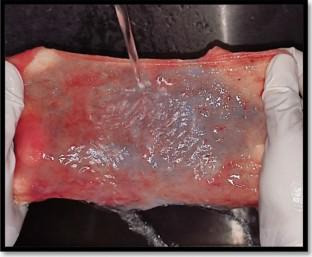
The purpose of the present study was to process and assess the effect of hydrated amnion chorion membrane and dehydrated amnion chorion membrane on proliferation of periodontal ligament (PDL) fibroblast cells. The amnion chorion membrane (ACM) from placenta of 18 systemically healthy patients was obtained from the Department of Obstetrics and Gynaecology. They were processed as hydrated and dehydrated based on different processing methods. The Periodontal ligament cells were obtained from periodontal ligament of freshly extracted premolars of systemically healthy patients, due to orthodontic reasons. The PDL cells were further cultured in laboratory and were exposed to hydrated and dehydrated amnion chorion membrane. The MTT assay was performed to assess the proliferation of PDL fibroblast cells after 24 and 48 h. The hydrated and dehydrated amnion chorion membrane showed proliferation of PDL fibroblasts after 24 and 48 h. The proliferation of PDL fibroblasts in hydrated (p = 0.043) and dehydrated (p = 0.050) amnion chorion membrane was statistically significant at the end of 24 and 48 h respectively. On inter-group comparison dehydrated ACM showed significant proliferation of PDL fibroblasts after 24 (p=0.014) and 48 h (p=0.019). Within the limits of the present study, it can be concluded: both hydrated and dehydrated amnion chorion membrane showed proliferationof PDL fibroblast cells. However, dehydrated ACM showed significant proliferation of PDL fibroblasts.
中文翻译:

水化和脱水处理羊膜绒毛膜对牙周膜成纤维细胞增殖的影响评价
本研究的目的是处理和评估水合羊膜绒毛膜和脱水羊膜绒毛膜对牙周膜(PDL)成纤维细胞增殖的影响。来自 18 名全身健康患者胎盘的羊膜绒毛膜 (ACM) 从妇产科获得。根据不同的加工方法将它们加工成水合和脱水的。由于正畸原因,牙周膜细胞是从全身健康患者新鲜拔除的前磨牙的牙周膜中获得的。PDL 细胞在实验室中进一步培养,并暴露于水合和脱水的羊膜绒毛膜。进行 MTT 测定以评估 24 小时和 48 小时后 PDL 成纤维细胞的增殖。水合和脱水的羊膜绒毛膜在 24 和 48 小时后显示出 PDL 成纤维细胞的增殖。PDL成纤维细胞在水合条件下的增殖(p = 0.043) 和脱水 ( p = 0.050) 羊膜绒毛膜分别在 24 小时和 48 小时结束时具有统计学意义。在组间比较中,脱水 ACM 在 24 小时 ( p =0.014) 和 48 小时 ( p =0.019) 后显示 PDL 成纤维细胞显着增殖。在本研究的范围内,可以得出结论:水合和脱水的羊膜绒毛膜均显示出PDL成纤维细胞的增殖。然而,脱水的 ACM 显示 PDL 成纤维细胞显着增殖。


 京公网安备 11010802027423号