Cell and Tissue Banking ( IF 1.5 ) Pub Date : 2023-03-20 , DOI: 10.1007/s10561-023-10084-2 Leila Taghiyar 1 , Hamideh Asadi 1, 2 , Mohamadreza Baghaban Eslaminejad 1
|
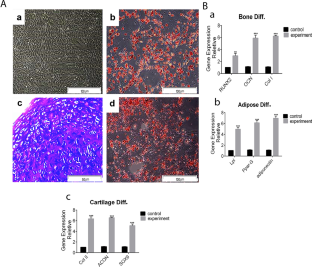
As a Natural decellularized extracellular matrix, osteochondral tissue is the best scaffold for the restoration of osteoarthritis defects. Bioscaffolds have the most similarly innate properties like biomechanical properties and the preserved connection of the bone-to-cartilage border. Although, their compacity and low porosity particularly, are proven to be difficulties of decellularization and cell penetration. This study aims to develop a new bioscaffold of decellularized osteochondral tissue (DOT) that is recellularized by bone marrow-derived mesenchymal stem cells (BM-MSCs), as a biphasic allograft, which preserved the interface between the cartilage section and subchondral bone of the joint. Whole osteochondral tissues of rabbit knee joints were sheeted in cartilaginous parts in 200–250 µm sections while connected to the subchondral bone and then fully decellularized. The BM-MSCs were seeded on the scaffolds in vitro; some constructs were subcutaneously implanted into the back of the rabbit. The cell penetration, differentiation to bone and cartilage, viability, and cell proliferation in vitro and in vivo were evaluated by qPCR, histological staining, MTT assay, and immunohistochemistry. DNA content analysis and SEM assessments confirmed the decellularization of the bioscaffold. Then, histological and SEM evaluations indicated that the cells could successfully penetrate the bone and cartilage lacunas in implanted grafts. MTT assay confirmed cell proliferation. Prominently, gene expression analysis showed that seeded cells differentiated into osteoblasts and chondrocytes in both bone and cartilage sections. More importantly, seeded cells on the bioscaffold started ECM secretion. Our results indicate that cartilage-to-bone border integrity was largely preserved. Additionally, ECM-sheeted DOT could be employed as a useful scaffold for promoting the regeneration of osteochondral defects.
中文翻译:
脱细胞全骨软骨片生物支架可改善兔模型中负载间充质干细胞的增殖和分化
骨软骨组织作为天然的脱细胞细胞外基质,是骨关节炎缺损修复的最佳支架。生物支架具有最相似的先天特性,如生物力学特性和骨与软骨边界的保留连接。然而,它们的致密性和低孔隙率被证明是脱细胞和细胞渗透的困难。本研究旨在开发一种新的脱细胞骨软骨组织(DOT)生物支架,该生物支架由骨髓来源的间充质干细胞(BM-MSC)再细胞化,作为双相同种异体移植物,保留了软骨部分和软骨下骨之间的界面。联合的。将兔膝关节的整个骨软骨组织切成200-250μm的软骨部分片,同时与软骨下骨连接,然后完全脱细胞。将BM-MSCs体外接种到支架上;一些构建体被皮下植入兔子的背部。通过 qPCR、组织学染色、MTT 测定和免疫组织化学评估体外和体内的细胞渗透、分化为骨和软骨、活力和细胞增殖。DNA 含量分析和 SEM 评估证实了生物支架的脱细胞化。然后,组织学和扫描电镜评估表明,细胞可以成功穿透植入移植物中的骨和软骨腔隙。MTT法证实细胞增殖。值得注意的是,基因表达分析表明,在骨和软骨切片中,接种的细胞分化为成骨细胞和软骨细胞。更重要的是,生物支架上的种子细胞开始分泌ECM。我们的结果表明软骨到骨边界的完整性在很大程度上得到了保留。此外,ECM片状DOT可用作促进骨软骨缺损再生的有用支架。


 京公网安备 11010802027423号