Journal of Molecular Histology ( IF 3.2 ) Pub Date : 2023-05-18 , DOI: 10.1007/s10735-023-10128-7 Gerardo Santamaría 1 , Aura Caterine Rengifo 1 , Orlando Torres-Fernández 1
|
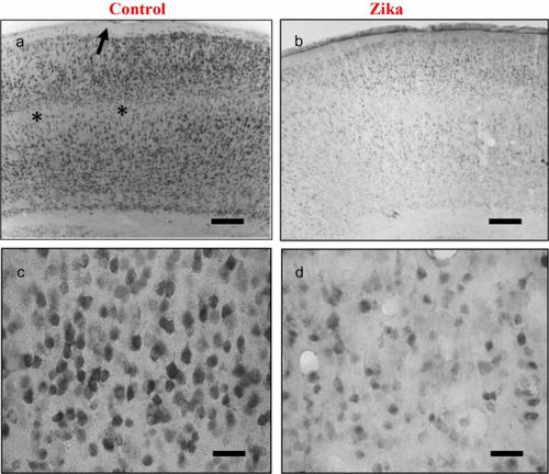
Microcephaly is the more severe brain malformation because of Zika virus infection. Increased vulnerability of neural stem and progenitor cells to Zika infection during prenatal neurodevelopment impairs the complete formation of cortical layers. Normal development of cerebellum is also affected. However, the follow-up of apparently healthy children born to Zika exposed mothers during pregnancy has revealed other neurological sequelae. This suggests Zika infection susceptibility remains in nervous tissue after neurogenesis end, when differentiated neuronal populations predominate. The neuronal nuclear protein (NeuN) is an exclusive marker of postmitotic neurons. Changes in NeuN expression are associated with neuronal degeneration. We have evaluated immunohistochemical expression of NeuN protein in cerebral cortex, hippocampus, and cerebellum of normal and Zika-infected neonatal Balb/c mice. The highest NeuN immunoreactivity was found mainly in neurons of all cortical layers, pyramidal layer of hippocampus, granular layer of dentate gyrus and in internal granular layer of cerebellum. Viral infection caused marked loss of NeuN immunostaining in all these brain areas. This suggests neurodegenerative effects of Zika virus infection during postmitotic neuron maturation and contribute to interpretation of neuropathogenic mechanisms of Zika.
中文翻译:
正常和寨卡感染乳鼠脑结构中的 NeuN 分布
小头畸形是由寨卡病毒感染引起的更严重的脑部畸形。在产前神经发育过程中,神经干细胞和祖细胞对寨卡病毒感染的脆弱性增加,损害了皮质层的完整形成。小脑的正常发育也会受到影响。然而,对感染寨卡病毒的母亲在怀孕期间所生的表面健康的孩子进行的随访发现了其他神经系统后遗症。这表明在神经发生结束后,当分化的神经元群体占主导地位时,寨卡病毒感染的易感性仍然存在于神经组织中。神经元核蛋白(NeuN)是有丝分裂后神经元的独特标记。NeuN 表达的变化与神经元变性有关。我们评估了大脑皮层、海马、以及正常和寨卡病毒感染的新生 Balb/c 小鼠的小脑。NeuN 免疫反应性最高的区域主要是所有皮质层、海马锥体层、齿状回颗粒层和小脑内颗粒层的神经元。病毒感染导致所有这些大脑区域的 NeuN 免疫染色显着丧失。这表明寨卡病毒感染在有丝分裂后神经元成熟过程中产生神经退行性影响,并有助于解释寨卡病毒的神经病理机制。


 京公网安备 11010802027423号