Journal of Biosciences ( IF 2.9 ) Pub Date : 2023-10-13 , DOI: 10.1007/s12038-023-00359-x Nazlar Ghasemzadeh , Fereidoun Nowshiravan Rahatabad , Siamak Haghipour , Shabnam Andalibi Miandoab , Keivan Maghooli
|
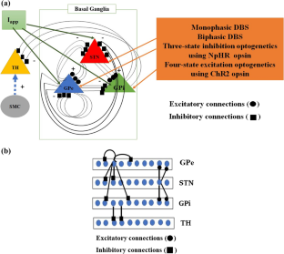
We examined electrical and optogenetic stimulations to explore their benefits and the effective range of their network effects. The error index (EI) and beta-activity of a network were considered by us as appropriate phenomena to compare stimulations. The basal ganglia (BG) network model considers areas of the brain affected by Parkinson’s disease (PD). It consists of the thalamus (TH), subthalamic nucleus (STN), globus pallidus interna (GPi,), and globus pallidus externa (GPe). To control the BG in PD, we stimulated the STN, GPe, and GPi with deep brain stimulation (DBS) and optogenetic stimulation, and to examine TH performance, we used the sensorimotor cortex (SMC). BG network performance was evaluated by measuring how TH responded to SMC input, and how STN, GPe, and Gpi were affected by DBS and optogenetic stimulation as evaluated using EI and beta-activity. By comparing the firing rates obtained from four applied stimulations at optimal conditions of the model, the effect of each stimulation was studied. The results indicated that applying monophasic DBS causes STN, GPi, and GPe cells to fire regularly, and biphasic DBS leads to increased oscillations between firings of STN cells; three-state optogenetic inhibition using halorhodopsin (NpHR) synchronizes the firing of GPe and GPi, and suppresses the neural activity of STN; and four-state optogenetic excitation using channelrhodopsin-2 (ChR2) incites the STN to excite and depolarize neural activity. All these events avoided pathological activation of PD and returned the performance to that of the healthy state. Finally, to evaluate our suggested method, we measured the beta-activity of monophasic and biphasic DBS and optogenetic inhibition and excitation by varying the \({\mathrm{electrical \ stimulation \ intensity }\ (A}_{elec})\) and optical stimulation intensity \(({A}_{light})\) and obtained optimal values for each state. According to our results, increasing \({A}_{elec}\) does not cause immediate decrease of beta-activity in monophasic and biphasic DBS. Increasing \({A}_{light}\) causes immediate decrease of beta-activity in NpHR, which then remains constant. In ChR2 there is no significant relation between beta-activity and \({A}_{light}.\)
中文翻译:
基于激发和抑制光遗传学模型以及单相和双相电刺激控制帕金森基底节的病理活动
我们检查了电刺激和光遗传学刺激,以探索它们的好处及其网络效应的有效范围。我们认为网络的误差指数 ( EI ) 和 beta 活动是比较刺激的适当现象。基底神经节 (BG) 网络模型考虑了受帕金森病 (PD) 影响的大脑区域。它由丘脑 (TH)、丘脑底核 (STN)、苍白球内核 (GPi) 和苍白球外核 (GPe) 组成。为了控制 PD 中的 BG,我们通过深部脑刺激 (DBS) 和光遗传学刺激刺激 STN、GPe 和 GPi,为了检查 TH 表现,我们使用感觉运动皮层 (SMC)。通过测量 TH 如何响应 SMC 输入以及 STN、GPe 和 Gpi 如何受到 DBS 和光遗传学刺激的影响(使用EI和 beta 活性进行评估)来评估 BG 网络性能。通过比较模型最佳条件下四次施加刺激所获得的放电率,研究了每次刺激的效果。结果表明,应用单相 DBS 会导致 STN、GPi 和 GPe 细胞定期放电,而双相 DBS 会导致 STN 细胞放电之间的振荡增加;使用盐视紫红质 (NpHR) 的三态光遗传学抑制可同步 GPe 和 GPi 的放电,并抑制 STN 的神经活动;使用视紫红质通道蛋白 2 (ChR2) 的四态光遗传学激发刺激 STN 来激发和去极化神经活动。所有这些事件都避免了帕金森病的病理激活,并使性能恢复到健康状态。最后,为了评估我们建议的方法,我们通过改变\({\mathrm{电\刺激\强度}\(A}_{elec})\)来测量单相和双相DBS的β活性以及光遗传学抑制和激发和光刺激强度\(({A}_{light})\)并获得每个状态的最佳值。根据我们的结果,增加\({A}_{elec}\)不会导致单相和双相 DBS 中 β 活性立即降低。增加\({A}_{light}\)会导致 NpHR 中的 β 活性立即降低,然后保持恒定。在 ChR2 中,β 活性和\({A}_{light}.\)之间没有显着关系


 京公网安备 11010802027423号