Journal of Neural Transmission ( IF 3.3 ) Pub Date : 2023-11-30 , DOI: 10.1007/s00702-023-02721-7 Giovanni Palermo 1 , Alessandro Galgani 2 , Gabriele Bellini 1 , Francesco Lombardo 3 , Nicola Martini 4 , Riccardo Morganti 5 , Davide Paoli 1 , Sara De Cori 3 , Francesca Frijia 4, 6 , Gabriele Siciliano 1 , Roberto Ceravolo 1 , Filippo Sean Giorgi 2
|
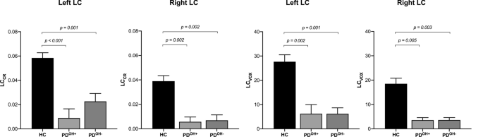
Locus coeruleus (LC) is the main noradrenergic nucleus of the brain, and degenerates early in Parkinson’s disease (PD). The objective of this study is to test whether degeneration of the LC is associated with orthostatic hypotension (OH) in PD. A total of 22 cognitively intact PD patients and 52 age-matched healthy volunteers underwent 3 T magnetic resonance (MRI) with neuromelanin-sensitive T1-weighted sequences (LC-MRI). For each subject, a template space-based LC-MRI was used to calculate LC signal intensity (LC contrast ratio—LCCR) and the estimated number of voxels (LCVOX) belonging to LC. Then, we compared the LC-MRI parameters in PD patients with OH (PDOH+) versus without OH (PDOH−) (matched for sex, age, and disease duration) using one-way analysis of variance followed by multiple comparison tests. We also tested for correlations between subject’s LC-MRI features and orthostatic drop in systolic blood pressure (SBP). PDOH− and PDOH+ did not differ significantly (p > 0.05) based on demographics and clinical characteristics, except for blood pressure measurements and SCOPA-AUT cardiovascular domain (p < 0.05). LCCR and LCVOX measures were significantly lower in PD compared to HC, while no differences were observed between PDOH− and PDOH+. Additionally, no correlation was found between the LC-MRI parameters and the orthostatic drop in SBP or the clinical severity of autonomic symptoms (p > 0.05). Conversely, RBD symptom severity negatively correlated with several LC-MRI parameters. Our results failed to indicate a link between the LC-MRI features and the presence of OH in PD but confirmed a marked alteration of LC signal in PD patients.
中文翻译:
帕金森病的神经源性直立性低血压:蓝斑磁共振成像有作用吗?
蓝斑 (LC) 是大脑的主要去甲肾上腺素能核,在帕金森病 (PD) 早期退化。本研究的目的是测试 LC 变性是否与 PD 中的体位性低血压 (OH) 有关。总共 22 名认知完整的 PD 患者和 52 名年龄匹配的健康志愿者接受了 3 T 磁共振 (MRI) 和神经黑色素敏感 T1 加权序列 (LC-MRI)。对于每个受试者,使用基于模板空间的 LC-MRI 来计算 LC 信号强度(LC 对比度 - LC CR )和属于 LC的估计体素数量(LC VOX )。然后,我们使用单向方差分析和多重比较检验,比较了患有 OH 的 PD 患者 (PD OH+ ) 与不患有 OH (PD OH− ) 的 LC-MRI 参数(匹配性别、年龄和病程)。我们还测试了受试者的 LC-MRI 特征与收缩压 (SBP) 直立性下降之间的相关性。除了血压测量和 SCOPA-AUT 心血管领域 ( p < 0.05) 之外,根据人口统计和临床特征,PD OH−和PD OH+没有显着差异 ( p > 0.05)。与 HC 相比,PD 中的LC CR和 LC VOX测量值显着较低,而 PD OH−和 PD OH+之间未观察到差异。此外,未发现 LC-MRI 参数与 SBP 直立性下降或自主神经症状的临床严重程度之间存在相关性 ( p > 0.05)。相反,RBD 症状严重程度与多个 LC-MRI 参数呈负相关。我们的结果未能表明 LC-MRI 特征与 PD 中 OH 的存在之间存在联系,但证实了 PD 患者中 LC 信号的显着改变。


 京公网安备 11010802027423号