SN Comprehensive Clinical Medicine Pub Date : 2023-12-22 , DOI: 10.1007/s42399-023-01631-9 Mohamed Henia , Stefan Linsler , Walter J. Schulz-Schaeffer , Steffi Urbschat , Julia Becker-Kettern , Malvina Garner , Joachim Oertel , Ralf Ketter
|
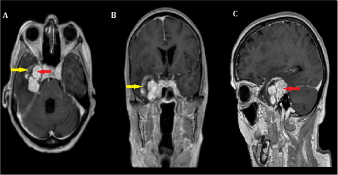
Intracranial schwannomas are relatively uncommon, accounting for approximately 8% of all intracranial tumors, while intracerebral schwannomas represent an even rarer entity, responsible for roughly 1% of all intracranial schwannomas. After reviewing the relevant literature, we discussed the clinical journey of a 74-year-old woman who presented with a 3-week history of dizziness and nausea. Magnetic resonance imaging revealed a right temporal mass lesion with perifocal edema. The initial suspicion was the diagnosis of a glioblastoma or metastasis, prompting surgical intervention. During the surgery, a gross total resection of a noninvasive tumor was successfully performed. The patient’s postoperative recovery was uneventful. Histopathological examination and confirmatory immunohistochemistry played a crucial role in reaching the final diagnosis of an intracerebral temporal schwannoma, highlighting the diagnostic challenges posed by radiologically indistinguishable features from metastasis and gliomas. Despite these challenges, complete surgical removal remains the most preferred treatment option, resulting in a favorable long-term prognosis without the need for adjuvant or neoadjuvant chemotherapy. Intracerebral schwannomas are exceedingly rare brain tumors, often found on the brain’s surface or adjacent ventricles. Early and accurate diagnosis can be challenging due to radiological features overlapping with other intracranial pathologies. Nonetheless, histopathological examination and immunohistochemistry remain indispensable tools in establishing a definitive diagnosis and guiding effective treatment strategies. With complete surgical excision, patients with intracerebral schwannomas can expect a positive outcome and a promising long-term prognosis. Further research and case studies are warranted to enhance our understanding of these rare tumors and improve patient outcomes.
中文翻译:
颅内脑神经鞘瘤:一例报告及文献复习
颅内神经鞘瘤相对罕见,约占所有颅内肿瘤的 8%,而脑内神经鞘瘤则更为罕见,约占所有颅内神经鞘瘤的 1%。在查阅相关文献后,我们讨论了一名 74 岁女性的临床经历,她有 3 周的头晕和恶心病史。磁共振成像显示右侧颞部肿块病变并伴有灶周水肿。最初的怀疑是胶质母细胞瘤或转移的诊断,促使进行手术干预。手术过程中,成功完成了非侵袭性肿瘤的大体全切除。患者术后恢复顺利。组织病理学检查和验证性免疫组织化学在脑内颞神经鞘瘤的最终诊断中发挥了至关重要的作用,突显了转移瘤和神经胶质瘤的放射学特征难以区分所带来的诊断挑战。尽管存在这些挑战,完全手术切除仍然是最优选的治疗选择,无需辅助或新辅助化疗即可获得良好的长期预后。脑内神经鞘瘤是极其罕见的脑肿瘤,通常发现于大脑表面或邻近的脑室。由于放射学特征与其他颅内病变重叠,早期和准确的诊断可能具有挑战性。尽管如此,组织病理学检查和免疫组织化学仍然是建立明确诊断和指导有效治疗策略不可或缺的工具。通过完全手术切除,脑内神经鞘瘤患者可以获得积极的结果和有希望的长期预后。需要进一步的研究和案例研究来增强我们对这些罕见肿瘤的了解并改善患者的治疗结果。


 京公网安备 11010802027423号