Biochemistry (Moscow) ( IF 2.8 ) Pub Date : 2024-02-14 , DOI: 10.1134/s0006297924010127 Anna O. Zholudeva , Nikolay S. Potapov , Ekaterina A. Kozlova , Maria E. Lomakina , Antonina Y. Alexandrova
|
Abstract
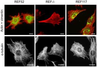
Cell migration is largely determined by the type of protrusions formed by the cell. Mesenchymal migration is accomplished by formation of lamellipodia and/or filopodia, while amoeboid migration is based on bleb formation. Changing of migrational conditions can lead to alteration in the character of cell movement. For example, inhibition of the Arp2/3-dependent actin polymerization by the CK-666 inhibitor leads to transition from mesenchymal to amoeboid motility mode. Ability of the cells to switch from one type of motility to another is called migratory plasticity. Cellular mechanisms regulating migratory plasticity are poorly understood. One of the factors determining the possibility of migratory plasticity may be the presence and/or organization of vimentin intermediate filaments (VIFs). To investigate whether organization of the VIF network affects the ability of fibroblasts to form membrane blebs, we used rat embryo fibroblasts REF52 with normal VIF organization, fibroblasts with vimentin knockout (REF–/–), and fibroblasts with mutation inhibiting assembly of the full-length VIFs (REF117). Blebs formation was induced by treatment of cells with CK-666. Vimentin knockout did not lead to statistically significant increase in the number of cells with blebs. The fibroblasts with short fragments of vimentin demonstrate the significant increase in number of cells forming blebs both spontaneously and in the presence of CK-666. Disruption of the VIF organization did not lead to the significant changes in the microtubules network or the level of myosin light chain phosphorylation, but caused significant reduction in the focal contact system. The most pronounced and statistically significant decrease in both size and number of focal adhesions were observed in the REF117 cells. We believe that regulation of the membrane blebbing by VIFs is mediated by their effect on the focal adhesion system. Analysis of migration of fibroblasts with different organization of VIFs in a three-dimensional collagen gel showed that organization of VIFs determines the type of cell protrusions, which, in turn, determines the character of cell movement. A novel role of VIFs as a regulator of membrane blebbing, essential for manifestation of the migratory plasticity, is shown.
中文翻译:
波形蛋白中间丝组装受损导致局灶接触的形成和成熟受到抑制以及细胞突起类型的改变
摘要
细胞迁移很大程度上取决于细胞形成的突起的类型。间充质迁移是通过形成片状伪足和/或丝状伪足来完成的,而变形虫迁移是基于泡形成。迁移条件的改变可以导致细胞运动特征的改变。例如,CK-666 抑制剂对 Arp2/3 依赖性肌动蛋白聚合的抑制导致从间充质运动模式转变为变形虫运动模式。细胞从一种运动类型转换为另一种运动类型的能力称为迁移可塑性。人们对调节迁移可塑性的细胞机制知之甚少。决定迁移可塑性可能性的因素之一可能是波形蛋白中间丝(VIF)的存在和/或组织。为了研究 VIF 网络的组织是否影响成纤维细胞形成膜泡的能力,我们使用了具有正常 VIF 组织的大鼠胚胎成纤维细胞 REF52、波形蛋白敲除的成纤维细胞 (REF –/– ) 以及具有抑制全膜组装的突变的成纤维细胞。长度 VIF (REF117)。通过用 CK-666 处理细胞来诱导气泡形成。波形蛋白敲除并没有导致带有泡的细胞数量出现统计上显着的增加。具有波形蛋白短片段的成纤维细胞表明自发形成泡的细胞数量以及在 CK-666 存在下均显着增加。 VIF组织的破坏并没有导致微管网络或肌球蛋白轻链磷酸化水平的显着变化,但引起局灶接触系统的显着减少。在 REF117 细胞中观察到粘着斑大小和数量最显着且具有统计学显着性的减少。我们认为,VIF 对膜起泡的调节是通过它们对粘着斑系统的影响来介导的。对三维胶原凝胶中具有不同 VIF 组织的成纤维细胞的迁移分析表明,VIF 的组织决定了细胞突起的类型,进而决定了细胞运动的特征。显示了 VIF 作为膜起泡调节剂的新作用,这对于表现迁移可塑性至关重要。


 京公网安备 11010802027423号