Cell and Tissue Research ( IF 3.6 ) Pub Date : 2024-03-15 , DOI: 10.1007/s00441-024-03883-w Shota Murase , Youhei Mantani , Nobuhiko Ohno , Asaka Shimada , Satoki Nakanishi , Rinako Morishita , Toshifumi Yokoyama , Nobuhiko Hoshi
|
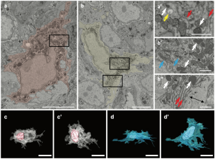
We previously clarified the histological characteristics of macrophages in the rat small intestine using serial block-face scanning electron microscopy (SBF-SEM). However, the regional differences in the characteristics of macrophages throughout the large intestine remain unknown. Here, we performed a pilot study to explore the regional differences in the ultrastructure of mucosal macrophages in the large intestine by using SBF-SEM analysis. SBF-SEM analysis conducted on the luminal side of the cecum and descending colon revealed macrophages as amorphous cells possessing abundant lysosomes and vacuoles. Macrophages in the cecum exhibited a higher abundance of lysosomes and a lower abundance of vacuoles than those in the descending colon. Macrophages with many intraepithelial cellular processes were observed beneath the intestinal superficial epithelium in the descending colon. Moreover, macrophages in contact with nerve fibers were more prevalent in the cecum than in the descending colon, and a subset of them surrounded a nerve bundle only in the cecum. In conclusion, the present pilot study suggested that the quantity of some organelles (lysosomes and vacuoles) in macrophages differed between the cecum and the descending colon and that there were some region-specific subsets of macrophages like nerve-associated macrophages in the cecum.
中文翻译:
大鼠大肠粘膜巨噬细胞超微结构的区域差异
我们之前使用连续块面扫描电子显微镜(SBF-SEM)阐明了大鼠小肠中巨噬细胞的组织学特征。然而,整个大肠巨噬细胞特征的区域差异仍然未知。在这里,我们进行了一项初步研究,利用 SBF-SEM 分析探讨大肠粘膜巨噬细胞超微结构的区域差异。对盲肠和降结肠管腔侧进行的 SBF-SEM 分析显示巨噬细胞是具有丰富溶酶体和液泡的无定形细胞。盲肠中的巨噬细胞比降结肠中的巨噬细胞表现出更高丰度的溶酶体和更低丰度的液泡。在降结肠肠浅表上皮下方观察到具有许多上皮内细胞突起的巨噬细胞。此外,与神经纤维接触的巨噬细胞在盲肠中比在降结肠中更普遍,并且其中的一部分仅在盲肠中包围神经束。总之,本初步研究表明,盲肠和降结肠中巨噬细胞中某些细胞器(溶酶体和液泡)的数量不同,并且盲肠中存在一些区域特异性的巨噬细胞亚群,例如神经相关巨噬细胞。


 京公网安备 11010802027423号